PEX5R rabbit pAb
- Catalog No.:YT7634
- Applications:WB;IHC
- Reactivity:Human;Mouse;Rat
- Target:
- PEX5R
- Fields:
- >>Peroxisome
- Gene Name:
- PEX5L PEX5R PXR2
- Protein Name:
- PEX5R
- Human Gene Id:
- 51555
- Human Swiss Prot No:
- Q8IYB4
- Mouse Gene Id:
- 58869
- Mouse Swiss Prot No:
- Q8C437
- Rat Gene Id:
- 286937
- Rat Swiss Prot No:
- Q925N3
- Immunogen:
- Synthesized peptide derived from human PEX5R AA range: 267-317
- Specificity:
- This antibody detects endogenous levels of PEX5R at Human/Mouse/Rat
- Formulation:
- Liquid in PBS containing 50% glycerol, 0.5% BSA and 0.02% sodium azide.
- Source:
- Polyclonal, Rabbit,IgG
- Dilution:
- WB 1:500-2000;IHC 1:50-300
- Purification:
- The antibody was affinity-purified from rabbit antiserum by affinity-chromatography using epitope-specific immunogen.
- Concentration:
- 1 mg/ml
- Storage Stability:
- -15°C to -25°C/1 year(Do not lower than -25°C)
- Molecular Weight(Da):
- 69kD
- Function:
- similarity:Belongs to the peroxisomal targeting signal receptor family.,similarity:Contains 5 TPR repeats.,subcellular location:Some fraction is membrane associated via its interaction with RAB8B.,subunit:Interacts with RAB8B (By similarity). May interact with the C-terminal PTS1-type tripeptide peroxisomal targeting signal (SKL-type); the relevance of such interaction is however unclear.,tissue specificity:Mainly expressed in brain. Also expressed in pancreas, testis and pituitary.,
- Subcellular Location:
- Cytoplasm . Membrane ; Peripheral membrane protein . Some fraction is membrane associated via its interaction with RAB8B. .
- Expression:
- Mainly expressed in brain. Also expressed in pancreas, testis and pituitary.
- June 19-2018
- WESTERN IMMUNOBLOTTING PROTOCOL
- June 19-2018
- IMMUNOHISTOCHEMISTRY-PARAFFIN PROTOCOL
- June 19-2018
- IMMUNOFLUORESCENCE PROTOCOL
- September 08-2020
- FLOW-CYTOMEYRT-PROTOCOL
- May 20-2022
- Cell-Based ELISA│解您多样本WB检测之困扰
- July 13-2018
- CELL-BASED-ELISA-PROTOCOL-FOR-ACETYL-PROTEIN
- July 13-2018
- CELL-BASED-ELISA-PROTOCOL-FOR-PHOSPHO-PROTEIN
- July 13-2018
- Antibody-FAQs
- Products Images
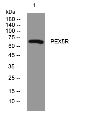
- Western blot analysis of lysates from Hela cells, primary antibody was diluted at 1:1000, 4°over night
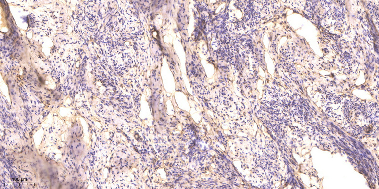
- Immunohistochemical analysis of paraffin-embedded human cervical carcinoma. 1, Antibody was diluted at 1:200(4° overnight). 2, Tris-EDTA,pH9.0 was used for antigen retrieval. 3,Secondary antibody was diluted at 1:200(room temperature, 45min).


