WIPF3 rabbit pAb
- Catalog No.:YT7587
- Applications:WB
- Reactivity:Human;Mouse;Rat
- Target:
- WIPF3
- Fields:
- >>Endocytosis;>>Pathogenic Escherichia coli infection;>>Yersinia infection
- Gene Name:
- WIPF3 CR16
- Protein Name:
- WIPF3
- Human Swiss Prot No:
- A6NGB9
- Mouse Swiss Prot No:
- P0C7L0
- Rat Gene Id:
- 259242
- Rat Swiss Prot No:
- Q9Z0G8
- Immunogen:
- Synthesized peptide derived from human WIPF3 AA range: 66-116
- Specificity:
- This antibody detects endogenous levels of WIPF3 at Human/Mouse/Rat
- Formulation:
- Liquid in PBS containing 50% glycerol, 0.5% BSA and 0.02% sodium azide.
- Source:
- Polyclonal, Rabbit,IgG
- Dilution:
- WB 1:500-2000
- Purification:
- The antibody was affinity-purified from rabbit antiserum by affinity-chromatography using epitope-specific immunogen.
- Concentration:
- 1 mg/ml
- Storage Stability:
- -15°C to -25°C/1 year(Do not lower than -25°C)
- Molecular Weight(Da):
- 53kD
- Function:
- domain:The KLKR motif is essential for G-actin binding and for actin polymerization.,domain:The profilin-binding motif has been implicated in the interaction with profilin and SH3 domains.,domain:The WH2 domain is found in a number of putative actin-binding proteins.,function:May be a regulator of cytoskeletal organization. May have a role in spermatogenesis.,similarity:Belongs to the verprolin family.,similarity:Contains 1 WH2 domain.,subcellular location:In hippocampal neurons colocalizes with WASL in the cell body, axons and the growth cone.,subunit:Interacts with WASL, and monomeric and filamentous actin.,
- Subcellular Location:
- Cytoplasm. In hippocampal neurons colocalizes with WASL in the cell body, axons and the growth cone. .
- June 19-2018
- WESTERN IMMUNOBLOTTING PROTOCOL
- June 19-2018
- IMMUNOHISTOCHEMISTRY-PARAFFIN PROTOCOL
- June 19-2018
- IMMUNOFLUORESCENCE PROTOCOL
- September 08-2020
- FLOW-CYTOMEYRT-PROTOCOL
- May 20-2022
- Cell-Based ELISA│解您多样本WB检测之困扰
- July 13-2018
- CELL-BASED-ELISA-PROTOCOL-FOR-ACETYL-PROTEIN
- July 13-2018
- CELL-BASED-ELISA-PROTOCOL-FOR-PHOSPHO-PROTEIN
- July 13-2018
- Antibody-FAQs
- Products Images
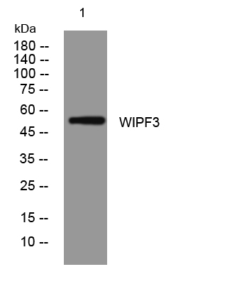
- Western blot analysis of lysates from A549 cells, primary antibody was diluted at 1:1000, 4°over night


