S36A2 rabbit pAb
- Catalog No.:YT7518
- Applications:WB
- Reactivity:Human;Mouse;Rat
- Target:
- S36A2
- Fields:
- >>Protein digestion and absorption
- Gene Name:
- SLC36A2 PAT2 TRAMD1
- Protein Name:
- S36A2
- Human Gene Id:
- 153201
- Human Swiss Prot No:
- Q495M3
- Mouse Gene Id:
- 246049
- Mouse Swiss Prot No:
- Q8BHK3
- Rat Gene Id:
- 246235
- Rat Swiss Prot No:
- Q8K415
- Immunogen:
- Synthesized peptide derived from human S36A2 AA range: 130-180
- Specificity:
- This antibody detects endogenous levels of S36A2 at Human/Mouse/Rat
- Formulation:
- Liquid in PBS containing 50% glycerol, 0.5% BSA and 0.02% sodium azide.
- Source:
- Polyclonal, Rabbit,IgG
- Dilution:
- WB 1:500-2000
- Purification:
- The antibody was affinity-purified from rabbit antiserum by affinity-chromatography using epitope-specific immunogen.
- Concentration:
- 1 mg/ml
- Storage Stability:
- -15°C to -25°C/1 year(Do not lower than -25°C)
- Molecular Weight(Da):
- 53kD
- Background:
- This gene encodes a pH-dependent proton-coupled amino acid transporter that belongs to the amino acid auxin permease 1 protein family. The encoded protein primarily transports small amino acids such as glycine, alanine and proline. Mutations in this gene are associated with iminoglycinuria and hyperglycinuria. [provided by RefSeq, Sep 2010],
- Function:
- function:Involved in a pH-dependent electrogenic neuronal transport and sequestration of small amino acids amino acids such as glycine, alanine and proline. Inhibited by sarcosine.,similarity:Belongs to the amino acid/polyamine transporter 2 family.,tissue specificity:Abundantly expressed in kidney and muscle.,
- Subcellular Location:
- Cell membrane ; Multi-pass membrane protein . Endoplasmic reticulum membrane . Recycling endosome membrane .
- Expression:
- Abundantly expressed in kidney and muscle. Expressed in the S1 segment of the proximal tubule close to the glomerulus.
- June 19-2018
- WESTERN IMMUNOBLOTTING PROTOCOL
- June 19-2018
- IMMUNOHISTOCHEMISTRY-PARAFFIN PROTOCOL
- June 19-2018
- IMMUNOFLUORESCENCE PROTOCOL
- September 08-2020
- FLOW-CYTOMEYRT-PROTOCOL
- May 20-2022
- Cell-Based ELISA│解您多样本WB检测之困扰
- July 13-2018
- CELL-BASED-ELISA-PROTOCOL-FOR-ACETYL-PROTEIN
- July 13-2018
- CELL-BASED-ELISA-PROTOCOL-FOR-PHOSPHO-PROTEIN
- July 13-2018
- Antibody-FAQs
- Products Images
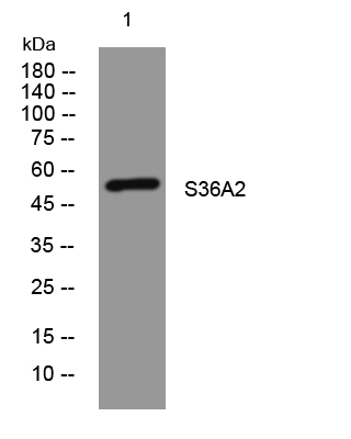
- Western blot analysis of lysates from 293T cells, primary antibody was diluted at 1:1000, 4°over night


