- Home
- About
- Promotions
-
Products
-
Elisa Kits
- |
-
Primary antibodies
- |
-
Secondary antibodies
- |
-
Proteins
- |
-
IHC reagents
- |
-
WB reagents
- PonceauS Staining Solution
- PBST Washing Buffer, 10X
- 1.5M Tris-HCl Buffer, pH8.8
- 1M Tris-HCl Buffer, pH6.8
- 10% SDS Solution
- Prestained Protein Marker
- TBST Washing Buffer, 10X
- SDS PAGE Loading Buffer, 5X
- Stripping Buffered Solution
- Tris Buffer, pH7.4, 10X
- Total Protein Extraction Kit
- Running Buffer, 10X
- Transfer Buffer, 10X
- 30% Acr-Bis(29:1) Solution
- Tris电泳液速溶颗粒
- PBS(1X, premixed powder)
- TBS(1X, premixed powder)
- 快速封闭液
- 转膜液速溶颗粒
- Chemical reagents
- News
- Distributor
- Resources
- Contact
- Home
- >
- Info
- >
- cGAS rabbit pAb
- >
- Go Back
cGAS rabbit pAb
- Catalog No.:YT7062
- Applications:WB
- Reactivity:Human;Mouse
- Fields:
- >>Cytosolic DNA-sensing pathway;>>Shigellosis;>>Human cytomegalovirus infection;>>Herpes simplex virus 1 infection;>>Human immunodeficiency virus 1 infection;>>Coronavirus disease - COVID-19
- Gene Name:
- MB21D1 cGAS C6orf150
- Immunogen:
- Synthesized peptide derived from human M21D1 AA range: 35-85
- Specificity:
- This antibody detects endogenous levels of M21D1 at Human
- Formulation:
- Liquid in PBS containing 50% glycerol, 0.5% BSA and 0.02% sodium azide.
- Source:
- Polyclonal, Rabbit,IgG
- Purification:
- The antibody was affinity-purified from rabbit antiserum by affinity-chromatography using epitope-specific immunogen.
- Storage Stability:
- -15°C to -25°C/1 year(Do not lower than -25°C)
- Molecular Weight(Da):
- 57kD
- Subcellular Location:
- Nucleus . Chromosome . Cell membrane ; Peripheral membrane protein . Cytoplasm, cytosol . Mainly localizes in the nucleus, and at low level in the cytosol (PubMed:31808743, PubMed:31544964). On chromosomes, enriched on centromeric satellite and LINE DNA repeat elements (PubMed:30811988). Exported from the nucleus to the cytosol in a XPO1/CRM1 via the nuclear export signal in response to DNA stimulation (PubMed:33406424). Outside the nucleus, localizes at the cell membrane as a peripheral membrane protein in resting conditions: association to the cell membrane is mediated via binding to phosphatidylinositol 4,5-bisphosphate (PtdIns(4,5)P2) (PubMed:30827685). Localization at the cell membrane is required to limit the recognition of self-DNA (PubMed:30827685). Following detection of double-st
- Expression:
- Expressed in the monocytic cell line THP1.
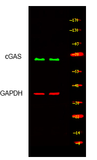
- Western blot analysis of lysates from MCF-7 and THP1 cells, primary antibody was diluted at 1:1000, 4°over night


