SDC3 rabbit pAb
- Catalog No.:YT6720
- Applications:WB;IHC
- Reactivity:Human;Mouse;Rat
- Target:
- SDC3
- Fields:
- >>Cell adhesion molecules
- Gene Name:
- SDC3 KIAA0468
- Protein Name:
- SDC3
- Human Gene Id:
- 9672
- Human Swiss Prot No:
- O75056
- Mouse Gene Id:
- 20970
- Mouse Swiss Prot No:
- Q64519
- Rat Gene Id:
- 116673
- Rat Swiss Prot No:
- P33671
- Immunogen:
- Synthesized peptide derived from human SDC3 AA range: 131-181
- Specificity:
- This antibody detects endogenous levels of SDC3 at Human/Mouse/Rat
- Formulation:
- Liquid in PBS containing 50% glycerol, 0.5% BSA and 0.02% sodium azide.
- Source:
- Polyclonal, Rabbit,IgG
- Dilution:
- WB 1:500-2000;IHC 1:50-300
- Purification:
- The antibody was affinity-purified from rabbit antiserum by affinity-chromatography using epitope-specific immunogen.
- Concentration:
- 1 mg/ml
- Storage Stability:
- -15°C to -25°C/1 year(Do not lower than -25°C)
- Molecular Weight(Da):
- 49kD
- Background:
- The protein encoded by this gene belongs to the syndecan proteoglycan family. It may play a role in the organization of cell shape by affecting the actin cytoskeleton, possibly by transferring signals from the cell surface in a sugar-dependent mechanism. Allelic variants of this gene have been associated with obesity. [provided by RefSeq, Oct 2009],
- Function:
- function:Cell surface proteoglycan that may bear heparan sulfate (By similarity). May have a role in the organization of cell shape by affecting the actin cytoskeleton, possibly by transferring signals from the cell surface in a sugar-dependent mechanism.,PTM:O-glycosylated within the Thr/Ser-rich region which could interact with lectin domains on other molecules.,similarity:Belongs to the syndecan proteoglycan family.,tissue specificity:Expressed in the nervous system, the adrenal gland, and the spleen.,
- Subcellular Location:
- Cell membrane; Single-pass type I membrane protein.
- Expression:
- Expressed in the nervous system, the adrenal gland, and the spleen.
- June 19-2018
- WESTERN IMMUNOBLOTTING PROTOCOL
- June 19-2018
- IMMUNOHISTOCHEMISTRY-PARAFFIN PROTOCOL
- June 19-2018
- IMMUNOFLUORESCENCE PROTOCOL
- September 08-2020
- FLOW-CYTOMEYRT-PROTOCOL
- May 20-2022
- Cell-Based ELISA│解您多样本WB检测之困扰
- July 13-2018
- CELL-BASED-ELISA-PROTOCOL-FOR-ACETYL-PROTEIN
- July 13-2018
- CELL-BASED-ELISA-PROTOCOL-FOR-PHOSPHO-PROTEIN
- July 13-2018
- Antibody-FAQs
- Products Images
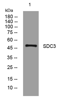
- Western blot analysis of lysates from HpeG2 cells, primary antibody was diluted at 1:1000, 4°over night
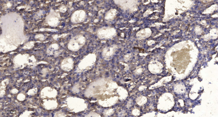
- Immunohistochemical analysis of paraffin-embedded human liver cancer. 1, Antibody was diluted at 1:200(4° overnight). 2, Tris-EDTA,pH9.0 was used for antigen retrieval. 3,Secondary antibody was diluted at 1:200(room temperature, 45min).


