- Home
- About
- Promotions
-
Products
-
Elisa Kits
- |
-
Primary antibodies
- |
-
Secondary antibodies
- |
-
Proteins
- |
-
IHC reagents
- |
-
WB reagents
- PonceauS Staining Solution
- PBST Washing Buffer, 10X
- 1.5M Tris-HCl Buffer, pH8.8
- 1M Tris-HCl Buffer, pH6.8
- 10% SDS Solution
- Prestained Protein Marker
- TBST Washing Buffer, 10X
- SDS PAGE Loading Buffer, 5X
- Stripping Buffered Solution
- Tris Buffer, pH7.4, 10X
- Total Protein Extraction Kit
- Running Buffer, 10X
- Transfer Buffer, 10X
- 30% Acr-Bis(29:1) Solution
- Tris电泳液速溶颗粒
- PBS(1X, premixed powder)
- TBS(1X, premixed powder)
- 快速封闭液
- 转膜液速溶颗粒
- Chemical reagents
- News
- Distributor
- Resources
- Contact
- Home
- >
- Info
- >
- CAR16 rabbit pAb
- >
- Go Back
CAR16 rabbit pAb
- Catalog No.:YT6533
- Applications:WB
- Reactivity:Human
- Fields:
- >>NOD-like receptor signaling pathway
- Gene Name:
- CARD16 COP COP1
- Immunogen:
- Synthesized peptide derived from human CAR16 AA range: 101-151
- Specificity:
- This antibody detects endogenous levels of CAR16 at Human
- Formulation:
- Liquid in PBS containing 50% glycerol, 0.5% BSA and 0.02% sodium azide.
- Source:
- Polyclonal, Rabbit,IgG
- Purification:
- The antibody was affinity-purified from rabbit antiserum by affinity-chromatography using epitope-specific immunogen.
- Storage Stability:
- -15°C to -25°C/1 year(Do not lower than -25°C)
- Molecular Weight(Da):
- 22kD
- Function:
- function:Caspase inhibitor. Acts as a regulator of procaspase-1/CASP1 activation implicated in the regulation of the proteolytic maturation of pro-interleukin-1 beta (IL1B) and its release during inflammation. Inhibits the release of IL1B in response to LPS in monocytes. Also induces NF-kappa-B activation during the pro-inflammatory cytokine response. Also able to inhibit CASP1-mediated neuronal cell death, TNF-alpha, hypoxia-, UV-, and staurosporine-mediated cell death but not ER stress-mediated cell death. Acts by preventing activation of caspases CASP1 and CASP4, possibly by preventing the interaction between CASP1 and RIPK2.,induction:Down-regulated in patients suffering of Huntington disease.,similarity:Contains 1 CARD domain.,subunit:Homooligomer. Interacts with CASP1, CASP4, CARD8 and RIPK2.,tissue specificity:Widely expressed. Expressed at higher level in placenta, spleen, lymph
- Expression:
- Widely expressed. Expressed at higher level in placenta, spleen, lymph node and bone marrow. Weakly or not expressed in thymus.
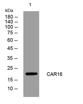
- Western blot analysis of lysates from Hela cells, primary antibody was diluted at 1:1000, 4°over night


