LRC26 rabbit pAb
- Catalog No.:YT6420
- Applications:WB;IHC
- Reactivity:Human;Mouse;Rat
- Target:
- LRC26
- Gene Name:
- LRRC26 CAPC
- Protein Name:
- LRC26
- Human Gene Id:
- 389816
- Human Swiss Prot No:
- Q2I0M4
- Mouse Gene Id:
- 227618
- Mouse Swiss Prot No:
- Q91W20
- Rat Gene Id:
- 311803
- Rat Swiss Prot No:
- Q6P7C4
- Immunogen:
- Synthesized peptide derived from human LRC26 AA range: 163-213
- Specificity:
- This antibody detects endogenous levels of LRC26 at Human/Mouse/Rat
- Formulation:
- Liquid in PBS containing 50% glycerol, 0.5% BSA and 0.02% sodium azide.
- Source:
- Polyclonal, Rabbit,IgG
- Dilution:
- WB 1:500-2000;IHC 1:50-300
- Purification:
- The antibody was affinity-purified from rabbit antiserum by affinity-chromatography using epitope-specific immunogen.
- Concentration:
- 1 mg/ml
- Storage Stability:
- -15°C to -25°C/1 year(Do not lower than -25°C)
- Molecular Weight(Da):
- 37kD
- Function:
- similarity:Contains 6 LRR (leucine-rich) repeats.,subcellular location:Although predicted to be a single-pass type I membrane protein, this protein localizes to the cytoplasm when expressed at high levels (PubMed:15164053).,tissue specificity:Expressed highly in normal prostate and salivary gland, very weakly in colon, pancreas, and intestine, and not at all in other tissues. Expressed highly in many cancer cell lines and in breast cancer, pancreatic cancer and colon cancer.,
- Subcellular Location:
- Cell membrane; Single-pass type I membrane protein. Cytoplasm, cytoskeleton. Localizes to the cytoplasm when expressed at high levels. .
- Expression:
- Isoform 1 is expressed highly in normal prostate and salivary gland, very weakly in colon, pancreas, and intestine, and not at all in other tissues. Isoform 1 is expressed highly in many cancer cell lines and in breast cancer, pancreatic cancer and colon cancer. Isoform 2 is expressed in cancer cell lines.
- June 19-2018
- WESTERN IMMUNOBLOTTING PROTOCOL
- June 19-2018
- IMMUNOHISTOCHEMISTRY-PARAFFIN PROTOCOL
- June 19-2018
- IMMUNOFLUORESCENCE PROTOCOL
- September 08-2020
- FLOW-CYTOMEYRT-PROTOCOL
- May 20-2022
- Cell-Based ELISA│解您多样本WB检测之困扰
- July 13-2018
- CELL-BASED-ELISA-PROTOCOL-FOR-ACETYL-PROTEIN
- July 13-2018
- CELL-BASED-ELISA-PROTOCOL-FOR-PHOSPHO-PROTEIN
- July 13-2018
- Antibody-FAQs
- Products Images
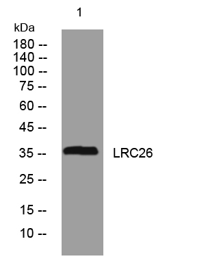
- Western blot analysis of lysates from SH-SY5Y cells, primary antibody was diluted at 1:1000, 4°over night
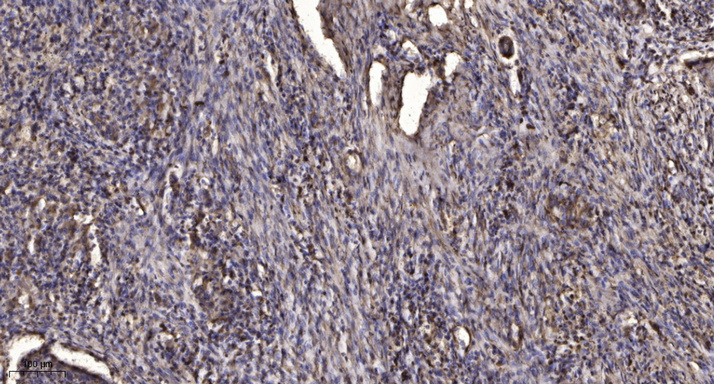
- Immunohistochemical analysis of paraffin-embedded human Squamous cell carcinoma of lung. 1, Antibody was diluted at 1:200(4° overnight). 2, Tris-EDTA,pH9.0 was used for antigen retrieval. 3,Secondary antibody was diluted at 1:200(room temperature, 45min).


