nNOS Polyclonal Antibody
- Catalog No.:YT3168
- Applications:WB;IHC;IF;ELISA
- Reactivity:Human;Mouse;Rat
- Target:
- NOS1/nNOS
- Fields:
- >>Arginine biosynthesis;>>Arginine and proline metabolism;>>Metabolic pathways;>>Calcium signaling pathway;>>Phagosome;>>Apelin signaling pathway;>>Circadian entrainment;>>Long-term depression;>>Relaxin signaling pathway;>>Salivary secretion;>>Alzheimer disease;>>Amyotrophic lateral sclerosis;>>Pathways of neurodegeneration - multiple diseases
- Gene Name:
- NOS1
- Protein Name:
- Nitric oxide synthase brain
- Human Gene Id:
- 4842
- Human Swiss Prot No:
- P29475
- Mouse Gene Id:
- 18125
- Mouse Swiss Prot No:
- Q9Z0J4
- Rat Gene Id:
- 24598
- Rat Swiss Prot No:
- P29476
- Immunogen:
- The antiserum was produced against synthesized peptide derived from human nNOS. AA range:818-867
- Specificity:
- NOS1 Polyclonal Antibody detects endogenous levels of NOS1 protein.
- Formulation:
- Liquid in PBS containing 50% glycerol, 0.5% BSA and 0.02% sodium azide.
- Source:
- Polyclonal, Rabbit,IgG
- Dilution:
- WB 1:500 - 1:2000. IHC 1:100 - 1:300. IF 1:200 - 1:1000. ELISA: 1:5000. Not yet tested in other applications.
- Purification:
- The antibody was affinity-purified from rabbit antiserum by affinity-chromatography using epitope-specific immunogen.
- Concentration:
- 1 mg/ml
- Storage Stability:
- -15°C to -25°C/1 year(Do not lower than -25°C)
- Other Name:
- NOS1;Nitric oxide synthase; brain;Constitutive NOS;NC-NOS;NOS type I;Neuronal NOS;N-NOS;nNOS;Peptidyl-cysteine S-nitrosylase NOS1;bNOS
- Observed Band(KD):
- 130-160kD
- Background:
- The protein encoded by this gene belongs to the family of nitric oxide synthases, which synthesize nitric oxide from L-arginine. Nitric oxide is a reactive free radical, which acts as a biologic mediator in several processes, including neurotransmission, and antimicrobial and antitumoral activities. In the brain and peripheral nervous system, nitric oxide displays many properties of a neurotransmitter, and has been implicated in neurotoxicity associated with stroke and neurodegenerative diseases, neural regulation of smooth muscle, including peristalsis, and penile erection. This protein is ubiquitously expressed, with high level of expression in skeletal muscle. Multiple transcript variants that differ in the 5' UTR have been described for this gene but the full-length nature of these transcripts is not known. Additionally, alternatively spliced transcript variants encoding different isoforms
- Function:
- alternative products:Isoform 3 is produced by different alternative splicing events implicating either the untranslated exons TEX1 (TN-NOS) or TEX1B (TN-NOSB) leading to a N-terminus truncated protein which possesses enzymatic activity comparable to that of isoform 1. The C-terminal truncated isoform 4 is produced by insertion of the TEX2 exon between exons 3 and 4 of isoform 1, leading to a frameshift and a premature stop codon,catalytic activity:L-arginine + n NADPH + n H(+) + m O(2) = citrulline + nitric oxide + n NADP(+).,cofactor:Binds 1 FAD.,cofactor:Binds 1 FMN.,cofactor:Heme group.,cofactor:Tetrahydrobiopterin (BH4). May stabilize the dimeric form of the enzyme.,disease:Genetic variations in NOS1 gene are associated with susceptibility to infantile hypertrophic pyloric stenosis type 1 (IHPS1) [MIM:179010]. IHPS has an incidence of 1-5 per 1'000 live births in whites and a marked
- Subcellular Location:
- Cell membrane, sarcolemma; Peripheral membrane protein. Cell projection, dendritic spine . In skeletal muscle, it is localized beneath the sarcolemma of fast-twitch muscle fiber by associating with the dystrophin glycoprotein complex. In neurons, enriched in dendritic spines (By similarity). .
- Expression:
- Isoform 1 is ubiquitously expressed: detected in skeletal muscle and brain, also in testis, lung and kidney, and at low levels in heart, adrenal gland and retina. Not detected in the platelets. Isoform 3 is expressed only in testis. Isoform 4 is detected in testis, skeletal muscle, lung, and kidney, at low levels in the brain, but not in the heart and adrenal gland.
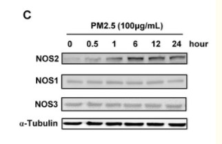
PM2.5 induces autophagy-mediated cell death via NOS2 signaling in human bronchial epithelium cells. International Journal of Biological Sciences Int J Biol Sci. 2018; 14(5): 557–564 WB Human BEASE-2B cell
Zhu, Xiao-Ming, et al. "PM2. 5 induces autophagy-mediated cell death via NOS2 signaling in human bronchial epithelium cells." International journal of biological sciences 14.5 (2018): 557.
- June 19-2018
- WESTERN IMMUNOBLOTTING PROTOCOL
- June 19-2018
- IMMUNOHISTOCHEMISTRY-PARAFFIN PROTOCOL
- June 19-2018
- IMMUNOFLUORESCENCE PROTOCOL
- September 08-2020
- FLOW-CYTOMEYRT-PROTOCOL
- May 20-2022
- Cell-Based ELISA│解您多样本WB检测之困扰
- July 13-2018
- CELL-BASED-ELISA-PROTOCOL-FOR-ACETYL-PROTEIN
- July 13-2018
- CELL-BASED-ELISA-PROTOCOL-FOR-PHOSPHO-PROTEIN
- July 13-2018
- Antibody-FAQs
- Products Images

- Zhu, Xiao-Ming, et al. "PM2. 5 induces autophagy-mediated cell death via NOS2 signaling in human bronchial epithelium cells." International journal of biological sciences 14.5 (2018): 557.

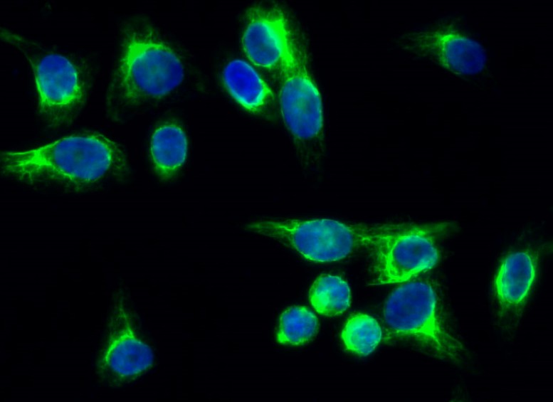
- Immunofluorescence analysis of Hela cell. 1,NOS1 Polyclonal Antibody(green) was diluted at 1:200(4° overnight). 2, Goat Anti Rabbit Alexa Fluor 488 Catalog:RS3211 was diluted at 1:1000(room temperature, 50min). 3 DAPI(blue) 10min.

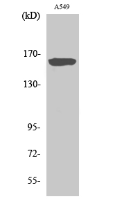
- Western Blot analysis of various cells using NOS1 Polyclonal Antibody diluted at 1:500

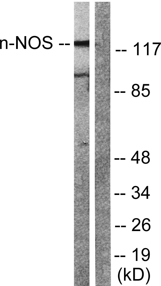
- Western blot analysis of lysates from Raw264.7 cells, treated with INF 2500u/ml 10', using nNOS Antibody. The lane on the right is blocked with the synthesized peptide.


