MMP-2 Polyclonal Antibody
- Catalog No.:YT2798
- Applications:WB;IHC;IF;ELISA
- Reactivity:Human;Mouse;Rat;Monkey
- Target:
- MMP-2
- Fields:
- >>Endocrine resistance;>>Leukocyte transendothelial migration;>>GnRH signaling pathway;>>Estrogen signaling pathway;>>Relaxin signaling pathway;>>AGE-RAGE signaling pathway in diabetic complications;>>Pathways in cancer;>>Proteoglycans in cancer;>>Bladder cancer;>>Diabetic cardiomyopathy;>>Fluid shear stress and atherosclerosis
- Gene Name:
- MMP2
- Protein Name:
- 72 kDa type IV collagenase
- Human Gene Id:
- 4313
- Human Swiss Prot No:
- P08253
- Mouse Gene Id:
- 17390
- Mouse Swiss Prot No:
- P33434
- Rat Gene Id:
- 81686
- Rat Swiss Prot No:
- P33436
- Immunogen:
- The antiserum was produced against synthesized peptide derived from human MMP-2. AA range:611-660
- Specificity:
- MMP-2 Polyclonal Antibody detects endogenous levels of MMP-2 protein.
- Formulation:
- Liquid in PBS containing 50% glycerol, 0.5% BSA and 0.02% sodium azide.
- Source:
- Polyclonal, Rabbit,IgG
- Dilution:
- WB 1:500 - 1:2000. IHC 1:100 - 1:300. IF 1:200 - 1:1000. ELISA: 1:20000. Not yet tested in other applications.
- Purification:
- The antibody was affinity-purified from rabbit antiserum by affinity-chromatography using epitope-specific immunogen.
- Concentration:
- 1 mg/ml
- Storage Stability:
- -15°C to -25°C/1 year(Do not lower than -25°C)
- Other Name:
- MMP2;CLG4A;72 kDa type IV collagenase;72 kDa gelatinase;Gelatinase A;Matrix metalloproteinase-2;MMP-2;TBE-1
- Observed Band(KD):
- 74kD
- Background:
- matrix metallopeptidase 2(MMP2) Homo sapiens This gene is a member of the matrix metalloproteinase (MMP) gene family, that are zinc-dependent enzymes capable of cleaving components of the extracellular matrix and molecules involved in signal transduction. The protein encoded by this gene is a gelatinase A, type IV collagenase, that contains three fibronectin type II repeats in its catalytic site that allow binding of denatured type IV and V collagen and elastin. Unlike most MMP family members, activation of this protein can occur on the cell membrane. This enzyme can be activated extracellularly by proteases, or, intracellulary by its S-glutathiolation with no requirement for proteolytical removal of the pro-domain. This protein is thought to be involved in multiple pathways including roles in the nervous system, endometrial menstrual breakdown, regulation of vascularization, and metastasis. Mutations in this gene have been associated with Win
- Function:
- catalytic activity:Cleavage of gelatin type I and collagen types IV, V, VII, X. Cleaves the collagen-like sequence Pro-Gln-Gly-|-Ile-Ala-Gly-Gln.,cofactor:Binds 2 zinc ions per subunit.,cofactor:Binds 4 calcium ions per subunit.,disease:Defects in MMP2 are the cause of Torg-Winchester syndrome [MIM:259600]; also called multicentric osteolysis nodulosis and arthropathy (MONA). Torg-Winchester syndrome is an autosomal recessive osteolysis syndrome. It is severe with generalized osteolysis and osteopenia. Subcutaneous nodules are usually absent. Torg-Winchester syndrome has been associated with a number of additional features including coarse face, corneal opacities, patches of thickened, hyperpigmented skin, hypertrichosis and gum hypertrophy. However, these features are not always present and have occasionally been observed in other osteolysis syndromes.,domain:The conserved cysteine pres
- Subcellular Location:
- [Isoform 1]: Secreted, extracellular space, extracellular matrix . Membrane. Nucleus. Colocalizes with integrin alphaV/beta3 at the membrane surface in angiogenic blood vessels and melanomas. Found in mitochondria, along microfibrils, and in nuclei of cardiomyocytes.; [Isoform 2]: Cytoplasm. Mitochondrion.
- Expression:
- Produced by normal skin fibroblasts. PEX is expressed in a number of tumors including gliomas, breast and prostate.
Methyl-CpG-binding domain 3 inhibits epithelial–mesenchymal transition in pancreatic cancer cells via TGF- β /Smad signalling. BRITISH JOURNAL OF CANCER 2016 Nov 29 WB Human SW1990 cell, PaTu8988 cell, PANC1 cell
LncRNA UCA1 promotes migration and invasion in pancreatic cancer cells via the Hippo pathway. BIOCHIMICA ET BIOPHYSICA ACTA-MOLECULAR BASIS OF DISEASE 2018 Mar 03 WB Human BxPC-3 cell, SW1990 cell,PaTu8988 cell, PANC-1 cell
GABAergic signaling facilitates breast cancer metastasis by promoting ERK1/2-dependent phosphorylation. CANCER LETTERS Cancer Lett. 2014 Jun;348:100 WB Human 1:1000 breast cancer tissues
MeCP2 suppresses LIN28A expression via binding to its methylated-CpG islands in pancreatic cancer cells. Oncotarget Oncotarget. 2016 Mar 22; 7(12): 14476–14485 WB Human BxPC3 cell, PANC1 cell, SW1990 cell,PaTu8988 cell
PHLDA2 regulates EMT and autophagy in colorectal cancer via the PI3K/AKT signaling pathway. Aging-US Aging-Us. 2020 May 15; 12(9): 7985–8000 WB Mouse 1:1000 HCT-116 cell-Xenograft , SW480 cell-Xenograft HCT-116 cell,SW480 cell
The role of SIRT1 in BMP2-induced chondrogenic differentiation and cartilage maintenance under oxidative stress.. Aging-US Aging-Us. 2020 May 31; 12(10): 9000–9013 WB Mouse 1:500 C3H10T1/2 cells
CBX4 promotes the proliferation and metastasis via regulating BMI‐1 in lung cancer. JOURNAL OF CELLULAR AND MOLECULAR MEDICINE J Cell Mol Med. 2020 Jan;24(1):618-631 WB Human 1:1000 NCI-H460 cell, A549 cell
Integration of exosomal miR-106a and mesothelial cells facilitates gastric cancer peritoneal dissemination. CELLULAR SIGNALLING Cell Signal. 2022 Mar;91:110230 WB Human 1:2000 HMrSV5 cell
Uridine-cytidine kinase 2 promotes metastasis of hepatocellular carcinoma cells via the Stat3 pathway. Cancer Management and Research Cancer Manag Res. 2018; 10: 6339–6355 WB Human 1:1000 Bel-7402 cell,Sk-hep-1 cell
Insulin-like growth factor II mRNA binding protein 3 regulates proliferation, invasion and migration of neuroendocrine cancer cells. International Journal of Clinical and Experimental Pathology Int J Clin Exp Patho. 2017; 10(10): 10269–10275 WB Mouse STC-1 cell
Protein arginine methyltransferase 1 coordinates the epithelial-mesenchymal transition/proliferation dichotomy in gastric cancer cells. EXPERIMENTAL CELL RESEARCH 2017 Oct 31 WB Human BGC823 cell, SGC7901 cell,AGS cell, MGC803 cell
YTH domain family 2 orchestrates epithelial-mesenchymal transition/proliferation dichotomy in pancreatic cancer cells. CELL CYCLE Cell Cycle. 2017;16(23):2259-2271 WB Human SW1990 cell, BxPC3 cell
Acute restraint stress triggers progesterone withdrawal and endometrial breakdown and shedding through corticosterone stimulation in mouse menstrual-like model. REPRODUCTION 2019 Feb 01 IHC Mouse 1:1500 endometrial tissues
Gene silencing of USP1 by lentivirus effectively inhibits proliferation and invasion of human osteosarcoma cells. INTERNATIONAL JOURNAL OF ONCOLOGY 2016 Dec 01 WB Human U2OS cell
Phosphoglycerate mutase 1 knockdown inhibits prostate cancer cell growth, migration, and invasion. ASIAN JOURNAL OF ANDROLOGY Asian J Androl. 2018 Mar-Apr; 20(2): 178–183 WB Human 1:500 PC-3 cell,22Rv1 cell,DU145 cell, LNCap cell
Long non‑coding RNA PVT1 promotes epithelial‑mesenchymal transition via the TGF‑β/Smad pathway in pancreatic cancer cells. ONCOLOGY REPORTS 2018 May 24 WB Human PaTu8988 cell, BxPc-3 cell
Endothelin-1 Activates the Notch Signaling Pathway and Promotes Tumorigenesis in Giant Cell Tumor of the Spine. SPINE Spine. 2019 Sep;44(17):E1000-E1009 WB Human 1:500 giant cell tumor (GCT) tissue
CCL5 secreted from bone marrow stromal cells stimulates the migration and invasion of Huh7 hepatocellular carcinoma cells via the PI3K-Akt pathway. INTERNATIONAL JOURNAL OF ONCOLOGY 2014 May 06 WB Human Huh7 cell
Oleuropein inhibits the proliferation and invasion of glioma cells via suppression of the AKT signaling pathway. ONCOLOGY REPORTS 2016 Jul 28 WB Human U251 cell, A172 cell
CTGF enhances resistance to 5-FU-mediating cell apoptosis through FAK/MEK/ERK signal pathway in colorectal cancer. OncoTargets and Therapy Oncotargets Ther. 2016; 9: 7285–7295 WB Human PaTu8988 cell, SW1990 cell
Emodin suppresses the migration and invasion of melanoma cells. BIOLOGICAL & PHARMACEUTICAL BULLETIN 2021 Mar 17 WB Mouse,Human 1 : 500 B16F10 Melanoma Cells, A375 Melanoma Cells
Paeonol exerts potential activities to inhibit the growth, migration and invasion of human gastric cancer BGC823 cells via downregulating MMP‑2 and MMP‑9. Molecular Medicine Reports Mol Med Rep. 2017 Nov;16(5):7513-7519 WB Human 1:1000 BGC823 cell
Effect of metformin on apoptosis, cell cycle arrest migration and invasion of A498 cells. Molecular Medicine Reports 2014 Mar 31 WB Human A498 cell
Expression of IMP3 as a marker for predicting poor outcome in gastroenteropancreatic neuroendocrine neoplasms. Oncology Letters 2017 Feb 14 IHC Human 1:100 gastroenteropancreatic neuroendocrine neoplasm (GEP‑NEN)
Fang, Zhiqing, et al. "Nitidine chloride inhibits renal cancer cell metastasis via suppressing AKT signaling pathway." Food and chemical toxicology 60 (2013): 246-251.
PHLDA2 regulates EMT and autophagy in colorectal cancer via the PI3K/AKT signaling pathway. Aging-US Aging-Us. 2020 May 15; 12(9): 7985–8000 WB Mouse 1:1000 HCT-116 cell-Xenograft , SW480 cell-Xenograft HCT-116 cell,SW480 cell
Nanocarrier of Pin1 inhibitor based on supercritical fluid technology inhibits cancer metastasis by blocking multiple signaling pathways Regenerative Biomaterials Dayun Yang WB,IHC Human,Mouse lung tissues HuH7 cell,HepG2 cell
LncRNA FAM83A-AS1 promotes epithelial–mesenchymal transition of pancreatic cancer cells via Hippo pathway. Min Xu WB Human BxPC-3 cell,PANC-1 cell, PaTu8988 cell
Rucaparib inhibits lung adenocarcinoma cell proliferation and migration via the SHCBP1/CDK1 pathway. FEBS Journal Rong Zhang WB Human 1 : 1000 A549 cell
MBD3 promotes epithelial-mesenchymal transition in gastric cancer cells by upregulating ACTG1 via the PI3K/AKT pathway BIOLOGICAL PROCEDURES ONLINE Wang Huizhi WB Human MGC-803 cell,HGC-27 cell
Nupr1-mediated vascular smooth muscle cell phenotype transformation involved in methamphetamine induces pulmonary hypertension CELL BIOLOGY AND TOXICOLOGY Zhou Jie WB Mouse pulmonary arterial smooth muscle cells (PASMCs)
Modulation of epithelial-mesenchymal transition by gemcitabine: Targeting ionizing radiation-induced cellular senescence in lung cancer cell BIOCHEMICAL PHARMACOLOGY Heng Zhou WB,IHC Mouse,Human 1:200 Lewis cell-Xenograft A549 cell
Blocking Sigmar1 exacerbates methamphetamine-induced hypertension BIOCHIMICA ET BIOPHYSICA ACTA-MOLECULAR BASIS OF DISEASE Zhen-Zhen Xu WB Mouse vascular smooth muscle cell (VSMC),C3H10T1/2 cell
Lnc-216 regulates the miR-143-5p /MMP2 signaling axis aggravates retinal endothelial cell dysfunction CLINICAL HEMORHEOLOGY AND MICROCIRCULATION Wang Fang WB Rat 1:1000 Rat retinal microvascular endothelial cell(RRMEC)
ATP11A Promotes Epithelial-mesenchymal Transition in Gastric Cancer Cells via the Hippo Pathway Journal of Cancer Wang Zhihua WB Human BGC-823 cell,MGC-803 cell,HGC-27 cell,SGC-7901 cell
Kaempferol inhibited invasion and metastasis of gastric cancer cells by targeting AKT/GSK3β pathway based on network pharmacology and molecular docking JOURNAL OF ASIAN NATURAL PRODUCTS RESEARCH Xia-Qing Gao WB Human 1:1000 HGC27 cell,MKN45 cell
- June 19-2018
- WESTERN IMMUNOBLOTTING PROTOCOL
- June 19-2018
- IMMUNOHISTOCHEMISTRY-PARAFFIN PROTOCOL
- June 19-2018
- IMMUNOFLUORESCENCE PROTOCOL
- September 08-2020
- FLOW-CYTOMEYRT-PROTOCOL
- May 20-2022
- Cell-Based ELISA│解您多样本WB检测之困扰
- July 13-2018
- CELL-BASED-ELISA-PROTOCOL-FOR-ACETYL-PROTEIN
- July 13-2018
- CELL-BASED-ELISA-PROTOCOL-FOR-PHOSPHO-PROTEIN
- July 13-2018
- Antibody-FAQs
- Products Images

- Rucaparib inhibits lung adenocarcinoma cell proliferation and migration via the SHCBP1/CDK1 pathway. FEBS Journal Rong Zhang WB Human 1 : 1000 A549 cell
.jpg)
- Wang, H., Li, J., He, J. et al. Methyl-CpG-binding protein 2 drives the Furin/TGF-β1/Smad axis to promote epithelial–mesenchymal transition in pancreatic cancer cells. Oncogenesis9, 76 (2020).
.jpg)
- Ma, Zhan, Shuping Lou, and Zheng Jiang. "PHLDA2 regulates EMT and autophagy in colorectal cancer via the PI3K/AKT signaling pathway." Aging (Albany NY) 12.9 (2020): 7985.
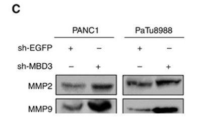
- Xu, Min, et al. "Methyl-CpG-binding domain 3 inhibits epithelial–mesenchymal transition in pancreatic cancer cells via TGF-β/Smad signalling." British journal of cancer 116.1 (2017): 91.
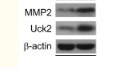
- Zhou, Qiming, et al. "Uridine-cytidine kinase 2 promotes metastasis of hepatocellular carcinoma cells via the Stat3 pathway." Cancer management and research 10 (2018): 6339.
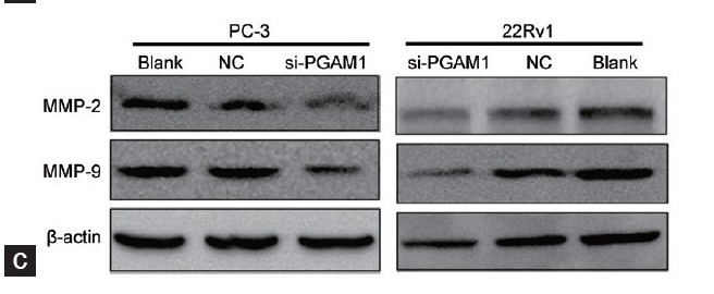
- Wen, Yao-An, et al. "Phosphoglycerate mutase 1 knockdown inhibits prostate cancer cell growth, migration, and invasion." Asian journal of andrology 20.2 (2018): 178.
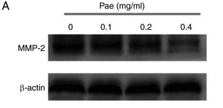
- Lyu, Zhong‑Kuan, et al. "Paeonol exerts potential activities to inhibit the growth, migration and invasion of human gastric cancer BGC823 cells via downregulating MMP‑2 and MMP‑9." Molecular medicine reports 16.5 (2017): 7513-7519.
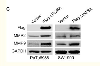
- Xu, Min, et al. "MeCP2 suppresses LIN28A expression via binding to its methylated-CpG islands in pancreatic cancer cells." Oncotarget 7.12 (2016): 14476.
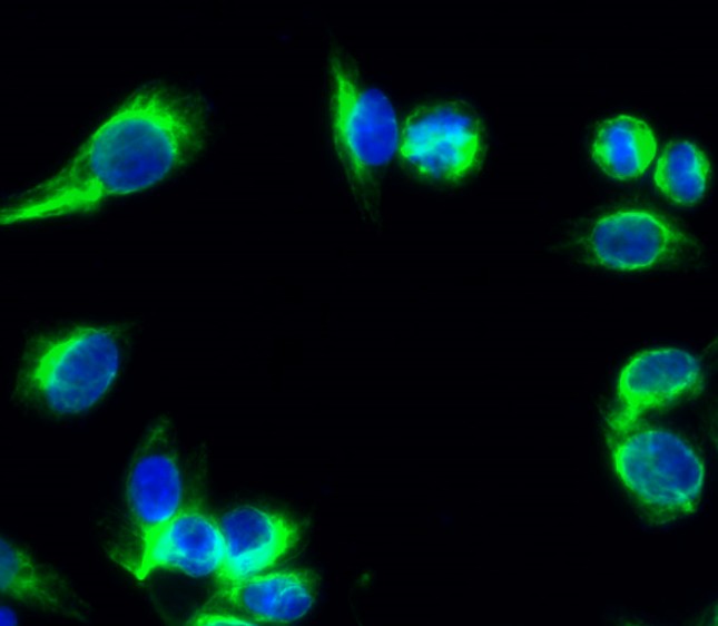
- Immunofluorescence analysis of Hela cell. 1,MMP-2 Polyclonal Antibody(green) was diluted at 1:200(4° overnight). 2, Goat Anti Rabbit Alexa Fluor 488 Catalog:RS3211 was diluted at 1:1000(room temperature, 50min). 3 DAPI(blue) 10min.
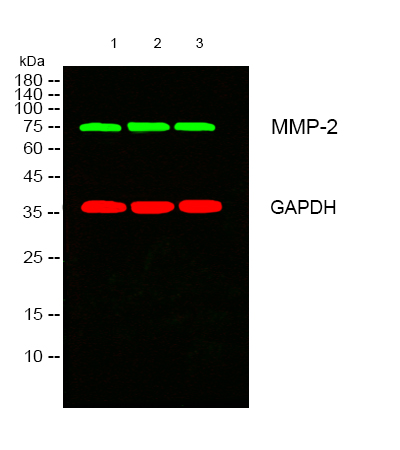
- Western blot analysis of lysates from 1) 3T3 , 2) Jurkat ,3) HT29 cells, (Green) primary antibody was diluted at 1:1000, 4°over night, secondary antibody(cat:RS23920)was diluted at 1:10000, 37° 1hour. (Red) GAPDH Monoclonal Antibody(2B8) (cat:YM3029) antibody was diluted at 1:5000 as loading control, 4° over night,secondary antibody(cat:RS23710)was diluted at 1:10000, 37° 1hour.
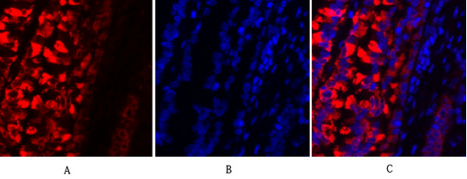
- Immunofluorescence analysis of rat-lung tissue. 1,MMP-2 Polyclonal Antibody(red) was diluted at 1:200(4°C,overnight). 2, Cy3 labled Secondary antibody was diluted at 1:300(room temperature, 50min).3, Picture B: DAPI(blue) 10min. Picture A:Target. Picture B: DAPI. Picture C: merge of A+B
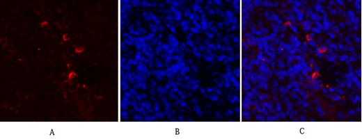
- Immunofluorescence analysis of rat-spleen tissue. 1,MMP-2 Polyclonal Antibody(red) was diluted at 1:200(4°C,overnight). 2, Cy3 labled Secondary antibody was diluted at 1:300(room temperature, 50min).3, Picture B: DAPI(blue) 10min. Picture A:Target. Picture B: DAPI. Picture C: merge of A+B
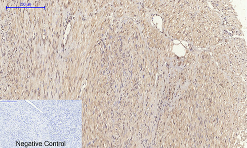
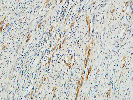
- Immunohistochemical analysis of paraffin-embedded Human-uterus-cancer tissue. 1,MMP-2 Polyclonal Antibody was diluted at 1:200(4°C,overnight). 2, Sodium citrate pH 6.0 was used for antibody retrieval(>98°C,20min). 3,Secondary antibody was diluted at 1:200(room tempeRature, 30min). Negative control was used by secondary antibody only.
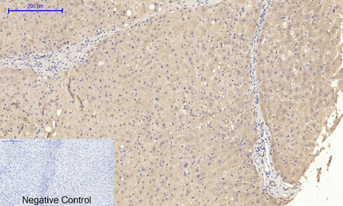
- Immunohistochemical analysis of paraffin-embedded Human-liver tissue. 1,MMP-2 Polyclonal Antibody was diluted at 1:200(4°C,overnight). 2, Sodium citrate pH 6.0 was used for antibody retrieval(>98°C,20min). 3,Secondary antibody was diluted at 1:200(room tempeRature, 30min). Negative control was used by secondary antibody only.
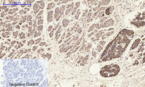
- Immunohistochemical analysis of paraffin-embedded Human-stomach-cancer tissue. 1,MMP-2 Polyclonal Antibody was diluted at 1:200(4°C,overnight). 2, Sodium citrate pH 6.0 was used for antibody retrieval(>98°C,20min). 3,Secondary antibody was diluted at 1:200(room tempeRature, 30min). Negative control was used by secondary antibody only.

- Immunohistochemical analysis of paraffin-embedded Rat-kidney tissue. 1,MMP-2 Polyclonal Antibody was diluted at 1:200(4°C,overnight). 2, Sodium citrate pH 6.0 was used for antibody retrieval(>98°C,20min). 3,Secondary antibody was diluted at 1:200(room tempeRature, 30min). Negative control was used by secondary antibody only.
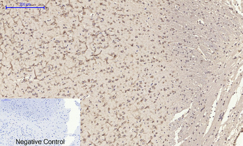
- Immunohistochemical analysis of paraffin-embedded Rat-brain tissue. 1,MMP-2 Polyclonal Antibody was diluted at 1:200(4°C,overnight). 2, Sodium citrate pH 6.0 was used for antibody retrieval(>98°C,20min). 3,Secondary antibody was diluted at 1:200(room tempeRature, 30min). Negative control was used by secondary antibody only.
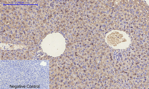
- Immunohistochemical analysis of paraffin-embedded Mouse-liver tissue. 1,MMP-2 Polyclonal Antibody was diluted at 1:200(4°C,overnight). 2, Sodium citrate pH 6.0 was used for antibody retrieval(>98°C,20min). 3,Secondary antibody was diluted at 1:200(room tempeRature, 30min). Negative control was used by secondary antibody only.
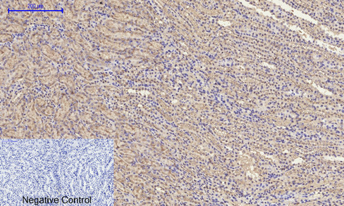
- Immunohistochemical analysis of paraffin-embedded Mouse-kidney tissue. 1,MMP-2 Polyclonal Antibody was diluted at 1:200(4°C,overnight). 2, Sodium citrate pH 6.0 was used for antibody retrieval(>98°C,20min). 3,Secondary antibody was diluted at 1:200(room tempeRature, 30min). Negative control was used by secondary antibody only.
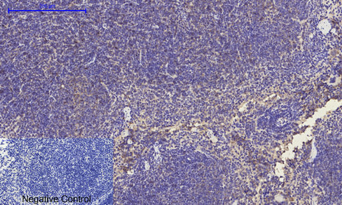
- Immunohistochemical analysis of paraffin-embedded Mouse-spleen tissue. 1,MMP-2 Polyclonal Antibody was diluted at 1:200(4°C,overnight). 2, Sodium citrate pH 6.0 was used for antibody retrieval(>98°C,20min). 3,Secondary antibody was diluted at 1:200(room tempeRature, 30min). Negative control was used by secondary antibody only.
- Immunohistochemical analysis of paraffin-embedded Human Endometrium. 1, Antibody was diluted at 1:200(4° overnight). 2, High-pressure and temperature EDTA, pH8.0 was used for antigen retrieval. 3,Secondary antibody was diluted at 1:200(room temperature, 30min).


