CAGE-1 Polyclonal Antibody
- Catalog No.:YT0606
- Applications:WB;IHC;IF;ELISA
- Reactivity:Human;Rat;Mouse;
- Target:
- CAGE-1
- Gene Name:
- CAGE1
- Protein Name:
- Cancer-associated gene 1 protein
- Human Gene Id:
- 285782
- Human Swiss Prot No:
- Q8TC20
- Mouse Swiss Prot No:
- Q5IR70
- Immunogen:
- The antiserum was produced against synthesized peptide derived from human CAGE1. AA range:711-760
- Specificity:
- CAGE-1 Polyclonal Antibody detects endogenous levels of CAGE-1 protein.
- Formulation:
- Liquid in PBS containing 50% glycerol, 0.5% BSA and 0.02% sodium azide.
- Source:
- Polyclonal, Rabbit,IgG
- Dilution:
- WB 1:500 - 1:2000. IHC 1:100 - 1:300. ELISA: 1:40000.. IF 1:50-200
- Purification:
- The antibody was affinity-purified from rabbit antiserum by affinity-chromatography using epitope-specific immunogen.
- Concentration:
- 1 mg/ml
- Storage Stability:
- -15°C to -25°C/1 year(Do not lower than -25°C)
- Other Name:
- CAGE1;CTAG3;Cancer-associated gene 1 protein;CAGE-1;Cancer/testis antigen 3;CT3
- Observed Band(KD):
- 90kD
- Background:
- tissue specificity:Testis-specific expression in normal tissues, but wide expression among cancer tissues and cell lines.,
- Function:
- tissue specificity:Testis-specific expression in normal tissues, but wide expression among cancer tissues and cell lines.,
- Expression:
- Testis-specific expression in normal tissues, but wide expression among cancer tissues and cell lines.
- June 19-2018
- WESTERN IMMUNOBLOTTING PROTOCOL
- June 19-2018
- IMMUNOHISTOCHEMISTRY-PARAFFIN PROTOCOL
- June 19-2018
- IMMUNOFLUORESCENCE PROTOCOL
- September 08-2020
- FLOW-CYTOMEYRT-PROTOCOL
- May 20-2022
- Cell-Based ELISA│解您多样本WB检测之困扰
- July 13-2018
- CELL-BASED-ELISA-PROTOCOL-FOR-ACETYL-PROTEIN
- July 13-2018
- CELL-BASED-ELISA-PROTOCOL-FOR-PHOSPHO-PROTEIN
- July 13-2018
- Antibody-FAQs
- Products Images
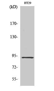
- Western Blot analysis of various cells using CAGE-1 Polyclonal Antibody diluted at 1:2000
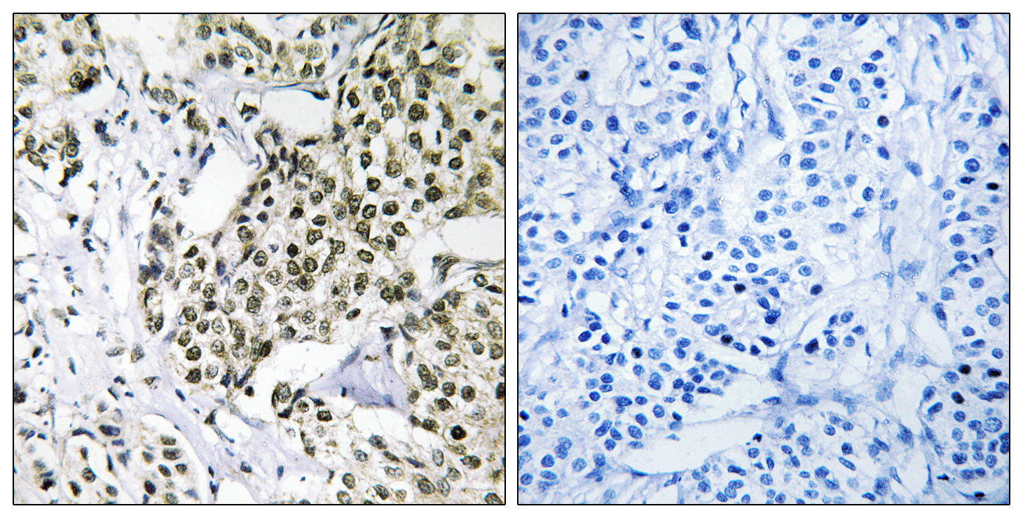
- Immunohistochemistry analysis of paraffin-embedded human breast carcinoma tissue, using CAGE1 Antibody. The picture on the right is blocked with the synthesized peptide.
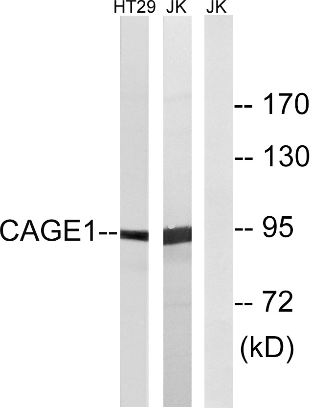
- Western blot analysis of lysates from HT-29 and Jurkat cells, using CAGE1 Antibody. The lane on the right is blocked with the synthesized peptide.

- Western blot analysis of the lysates from HepG2 cells using CAGE1 antibody.


