JNK1/2/3 (phospho Thr183/Y185) Polyclonal Antibody
- Catalog No.:YP0157
- Applications:IF;WB;IHC;ELISA
- Reactivity:Human;Mouse;Rat;Chicken/pig/fish(tested by our customer)
- Target:
- JNK1/2/3
- Fields:
- >>Endocrine resistance;>>MAPK signaling pathway;>>ErbB signaling pathway;>>Ras signaling pathway;>>cAMP signaling pathway;>>FoxO signaling pathway;>>Sphingolipid signaling pathway;>>Mitophagy - animal;>>Autophagy - animal;>>Protein processing in endoplasmic reticulum;>>Apoptosis;>>Apoptosis - multiple species;>>Necroptosis;>>Wnt signaling pathway;>>Osteoclast differentiation;>>Focal adhesion;>>Tight junction;>>Toll-like receptor signaling pathway;>>NOD-like receptor signaling pathway;>>RIG-I-like receptor signaling pathway;>>C-type lectin receptor signaling pathway;>>IL-17 signaling pathway;>>Th1 and Th2 cell differentiation;>>Th17 cell differentiation;>>T cell receptor signaling pathway;>>Fc epsilon RI signaling pathway;>>TNF signaling pathway;>>Neurotrophin signaling pathway;>>Retrograde endocannabinoid signaling;>>Dopaminergic synapse;>>Inflammatory mediator regulation of TRP channels;>>Insulin signaling pathway;>>GnRH signaling pathway;>>Progesterone-mediated oocyte maturation;>>Pr
- Gene Name:
- MAPK8/9/10
- Protein Name:
- Mitogen-activated protein kinase 8/9/10
- Human Gene Id:
- 5599/5601/5602
- Human Swiss Prot No:
- P45983/P45984/P53779
- Mouse Gene Id:
- 26419/26420
- Rat Gene Id:
- 116554/50658/25272
- Rat Swiss Prot No:
- P49185/P49186/P49187
- Immunogen:
- The antiserum was produced against synthesized peptide derived from human JNK1/2/3 around the phosphorylation site of Thr183 and Tyr185. AA range:151-200
- Specificity:
- Phospho-JNK1/2/3 (T183/Y185) Polyclonal Antibody detects endogenous levels of JNK1/2/3 protein only when phosphorylated at T183/Y185.
- Formulation:
- Liquid in PBS containing 50% glycerol, 0.5% BSA and 0.02% sodium azide.
- Source:
- Polyclonal, Rabbit,IgG
- Dilution:
- IF 1:50-200 WB 1:500-2000, IHC 1:50-300 IHC 1:50-300
- Purification:
- The antibody was affinity-purified from rabbit antiserum by affinity-chromatography using epitope-specific immunogen.
- Concentration:
- 1 mg/ml
- Storage Stability:
- -15°C to -25°C/1 year(Do not lower than -25°C)
- Other Name:
- MAPK8;JNK1;PRKM8;SAPK1;SAPK1C;Mitogen-activated protein kinase 8;MAP kinase 8;MAPK 8;JNK-46;Stress-activated protein kinase 1c;SAPK1c;Stress-activated protein kinase JNK1;c-Jun N-terminal kinase 1;MAPK9;JNK2;PRKM9;SAPK1A;Mi
- Observed Band(KD):
- 46kD,54kD
- Background:
- The protein encoded by this gene is a member of the MAP kinase family. MAP kinases act as an integration point for multiple biochemical signals, and are involved in a wide variety of cellular processes such as proliferation, differentiation, transcription regulation and development. This kinase is activated by various cell stimuli, and targets specific transcription factors, and thus mediates immediate-early gene expression in response to cell stimuli. The activation of this kinase by tumor-necrosis factor alpha (TNF-alpha) is found to be required for TNF-alpha induced apoptosis. This kinase is also involved in UV radiation induced apoptosis, which is thought to be related to cytochrom c-mediated cell death pathway. Studies of the mouse counterpart of this gene suggested that this kinase play a key role in T cell proliferation, apoptosis and differentiation. Several alternatively spl
- Function:
- catalytic activity:ATP + a protein = ADP + a phosphoprotein.,cofactor:Magnesium.,domain:The TXY motif contains the threonine and tyrosine residues whose phosphorylation activates the MAP kinases.,enzyme regulation:Activated by threonine and tyrosine phosphorylation by either of two dual specificity kinases, MAP2K4 and MAP2K7. Inhibited by dual specificity phosphatases, such as DUSP1.,function:JNK1 isoforms display different binding patterns: beta-1 preferentially binds to c-Jun, whereas alpha-1, alpha-2, and beta-2 have a similar low level of binding to both c-Jun or ATF2. However, there is no correlation between binding and phosphorylation, which is achieved at about the same efficiency by all isoforms.,function:Responds to activation by environmental stress and pro-inflammatory cytokines by phosphorylating a number of transcription factors, primarily components of AP-1 such as JUN, JDP
- Subcellular Location:
- Cytoplasm . Nucleus . Cell junction, synapse . In the cortical neurons, predominantly cytoplasmic and associated with the Golgi apparatus and endosomal fraction. Increased neuronal activity increases phosphorylated form at synapses (By similarity). Colocalizes with POU5F1 in the nucleus. .
- Expression:
- Brain,Epithelium,Fetal brain,Lung,Pooled,Testis,
NCX1 disturbs calcium homeostasis and promotes RANKL-induced osteoclast differentiation by regulating JNK/c-Fos/NFATc1 signaling pathway in multiple myeloma
IL-17A promotes endothelial cell senescence by up-regulating the expression of FTO through activating JNK signal pathway BIOGERONTOLOGY Li, Na, Luo, Runan, Zhang, Wenlong, Wu, Yu, Hu, Chaojie, Liu, Manli, Jiang, Diya, Jiang, Ziran, Zhao, Xinxin, Wang, Yiping, Li, Qing WB Human HUVECs
Therapeutic Potential of Synthetic Human β-Defensin 1 Short Motif Pep-B on Lipopolysaccharide-Stimulated Human Dental Pulp Stem Cells Mediat Inflamm. 2022;2022:6141967. WB Human 1 : 1000
Inhibitory effects of oxymatrine on TGF‑β1‑induced proliferation and abnormal differentiation in rat cardiac fibroblasts via the p38MAPK and ERK1/2 signaling pathways. Molecular Medicine Reports Mol Med Rep. 2017 Oct;16(4):5354-5362 WB Rat 1:1000 cardiac fibroblast (CFB)
Dibromoacetic Acid Induced Hepatotoxicity in Mice through Oxidative Stress and Toll-Like Receptor 4 Signaling Pathway Activation. Oxidative Medicine and Cellular Longevity Oxid Med Cell Longev. 2019;2019:5637235 WB Mouse Liver
Adipose-Derived Stem Cells Protect Ischemia-Reperfusion and Partial Hepatectomy by Attenuating Endoplasmic Reticulum Stress. Frontiers in Cell and Developmental Biology Front Cell Dev Biol. 2020 Mar;0:177 WB Pig Liver
Effect of conditioned medium from adipose derived mesenchymal stem cells on endoplasmic reticulum stress and lipid metabolism after hepatic ischemia reperfusion injury and hepatectomy in swine. LIFE SCIENCES Life Sci. 2021 Dec;:120212 WB Pig 1:1000 Liver
Administration of SB203580, a p38 MAPK Inhibitor, Reduced the Expression of MMP9, and Relieved Neurologic Severity in the Experimental Autoimmune Neuritis (EAN) in Rats. NEUROCHEMICAL RESEARCH Neurochem Res. 2015 Jul;40(7):1410-1420 WB Rat 1:1000 sciatic nerves
NOV inhibits proliferation while promoting apoptosis and migration in osteosarcoma cell lines through p38/MAPK and JNK/MAPK pathways. ONCOLOGY REPORTS 2015 Jul 24 WB Human 143B cell
17β-Estradiol on the Expression of G-Protein Coupled Estrogen Receptor (GPER/GPR30) Mitophagy, and the PI3K/Akt Signaling Pathway in ATDC5 Chondrocytes In Vitro. MEDICAL SCIENCE MONITOR Med Sci Monitor. 2018; 24: 1936–1947 WB Mouse ATDC5 cell
The negative charge of the 343 site is essential for maintaining physiological functions of CXCR4. BMC Molecular and Cell Biology Bmc Mol Cell Biol. 2021 Dec;22(1):1-10 WB Human 1:1000 HeLa cell
Sweroside promotes osteoblastic differentiation and mineralization via interaction of membrane estrogen receptor-α and GPR30 mediated p38 signalling pathway on MC3T3-E1 cells. PHYTOMEDICINE 2019 Dec 07 WB Human 1:1000 MC3T3-E1 cell
Effect of cold stress on the MAPK pathway and lipidomics on muscle of Takifugu fasciatus. AQUACULTURE Aquaculture. 2021 Jul;540:736691 WB Fish 1:1500 Muscles
β-Conglycinin-Induced Intestinal Porcine Epithelial Cell Damage via the Nuclear Factor κB/Mitogen-Activated Protein Kinase Signaling Pathway. JOURNAL OF AGRICULTURAL AND FOOD CHEMISTRY J Agr Food Chem. 2019;67(32):9009–9021 WB Pig 1:500 IPEC-J2 cell
Soybean Glycinin- and β-Conglycinin-Induced Intestinal Damage in Piglets via the p38/JNK/NF-κB Signaling Pathway. JOURNAL OF AGRICULTURAL AND FOOD CHEMISTRY J Agr Food Chem. 2018;66(36):9534–9541 WB Pig 1:1000 small intestine
Non‑canonical Wnt signaling contributes to ventilator‑induced lung injury through upregulation of WISP1 expression. INTERNATIONAL JOURNAL OF MOLECULAR MEDICINE 2019 Mar 01 IHC Mouse 1:200 lung
Lycium barbarum Polysaccharides Protect Rat Corneal Epithelial Cells against Ultraviolet B-Induced Apoptosis by Attenuating the Mitochondrial Pathway and Inhibiting JNK Phosphorylation. Biomed Research International Biomed Res Int. 2017;2017:5806832 WB Human,Rat 1 : 500 RCE cell
Protective effects of flavonoids from the leaves of Carya cathayensis Sarg. against H2O2‑induced oxidative damage and apoptosis in vitro. Experimental and Therapeutic Medicine Exp Ther Med. 2021 Dec;22(6):1-11 WB Rat 1:1000 Rat aortic endothelial cells (RAECs)
Ping-Chong-Jiang-Ni Formula Induces Apoptosis and Inhibits Proliferation of Human Ectopic Endometrial Stromal Cells in Endometriosis via the Activation of JNK Signaling Pathway. Evidence-based Complementary and Alternative Medicine Evid-Based Compl Alt. 2017;2017:6489427 WB Human 1 : 1000 EESCs
Icariin inhibits MMP‑1, MMP‑3 and MMP‑13 expression through MAPK pathways in IL‑1β‑stimulated SW1353 chondrosarcoma cells. Molecular Medicine Reports 2017 May 01 WB Human 1:1000 SW1353 cell
Pretreatment with pPolyHb attenuates H2O2-induced endothelial cell injury through inhibition of JNK/p38 MAPK pathway by upregulation of heme oxygenase-1. Artificial Cells Nanomedicine and Biotechnology Artif Cell Nanomed B. 2015;43(3):163-173 WB Human 1:500 HUVECs
Lu, Lin, et al. "MicroRNA-148b regulates tumor growth of non-small cell lung cancer through targeting MAPK/JNK pathway." BMC cancer 19.1 (2019): 209.
Peng, Chenglu, et al. "Glycinin-induced porcine IPEC-J2 cells damage via the NF-κB/MAPK signaling pathway." (2020).
Exosomes from adipose-derived mesenchymal stem cells can attenuate liver injury caused by minimally invasive hemihepatectomy combined with ischemia-reperfusion in minipigs by modulating the endoplasmic reticulum stress response LIFE SCIENCES Hongbin Wang WB Pig liver tissues adipose-derived mesenchymal stem cells (ADSCs)
Protective effect of cornuside on OGD/R injury in SH-SY5Y cells and its underlying mechanism. BRAIN RESEARCH Jianwei Gong WB Human SH-SY5Y cell
Design and synthesis of pterostilbene derivatives bearing triazole moiety that might treat DSS-induced colitis in mice through modulation of NF-κB/MAPK signaling pathways EUROPEAN JOURNAL OF MEDICINAL CHEMISTRY Liuzeng Chen WB Mouse 1:1000 RAW264.7 cell
Design, synthesis, in vitro and in vivo biological evaluation of pterostilbene derivatives for anti-inflammation therapy JOURNAL OF ENZYME INHIBITION AND MEDICINAL CHEMISTRY Liuzeng Chen WB Mouse 1:1000 RAW264.7 cell
Silencing miR-155-5p Alleviates Hippocampal Damage in Kainic Acid-Induced Epileptic Rats via the Dusp14/MAPK Pathway BRAIN RESEARCH BULLETIN Qiong Fang WB Rat 1:1000 hippocampal tissue
- June 19-2018
- WESTERN IMMUNOBLOTTING PROTOCOL
- June 19-2018
- IMMUNOHISTOCHEMISTRY-PARAFFIN PROTOCOL
- June 19-2018
- IMMUNOFLUORESCENCE PROTOCOL
- September 08-2020
- FLOW-CYTOMEYRT-PROTOCOL
- May 20-2022
- Cell-Based ELISA│解您多样本WB检测之困扰
- July 13-2018
- CELL-BASED-ELISA-PROTOCOL-FOR-ACETYL-PROTEIN
- July 13-2018
- CELL-BASED-ELISA-PROTOCOL-FOR-PHOSPHO-PROTEIN
- July 13-2018
- Antibody-FAQs
- Products Images
- Xu, Yini, et al. "Inhibitory effects of oxymatrine on TGF‑β1‑induced proliferation and abnormal differentiation in rat cardiac fibroblasts via the p38MAPK and ERK1/2 signaling pathways." Molecular medicine reports 16.4 (2017): 5354-5362.
-if-66.jpg)
- Immunofluorescence analysis of rat-liver tissue. 1,JNK1/2/3 (phospho Thr183/Y185) Polyclonal Antibody(red) was diluted at 1:200(4°C,overnight). 2, Cy3 labled Secondary antibody was diluted at 1:300(room temperature, 50min).3, Picture B: DAPI(blue) 10min. Picture A:Target. Picture B: DAPI. Picture C: merge of A+B
-if-67.jpg)
- Immunofluorescence analysis of rat-liver tissue. 1,JNK1/2/3 (phospho Thr183/Y185) Polyclonal Antibody(red) was diluted at 1:200(4°C,overnight). 2, Cy3 labled Secondary antibody was diluted at 1:300(room temperature, 50min).3, Picture B: DAPI(blue) 10min. Picture A:Target. Picture B: DAPI. Picture C: merge of A+B
-if-68.jpg)
- Immunofluorescence analysis of rat-spleen tissue. 1,JNK1/2/3 (phospho Thr183/Y185) Polyclonal Antibody(red) was diluted at 1:200(4°C,overnight). 2, Cy3 labled Secondary antibody was diluted at 1:300(room temperature, 50min).3, Picture B: DAPI(blue) 10min. Picture A:Target. Picture B: DAPI. Picture C: merge of A+B
-if-69.jpg)
- Immunofluorescence analysis of rat-spleen tissue. 1,JNK1/2/3 (phospho Thr183/Y185) Polyclonal Antibody(red) was diluted at 1:200(4°C,overnight). 2, Cy3 labled Secondary antibody was diluted at 1:300(room temperature, 50min).3, Picture B: DAPI(blue) 10min. Picture A:Target. Picture B: DAPI. Picture C: merge of A+B
-if-70.jpg)
- Immunofluorescence analysis of mouse-liver tissue. 1,JNK1/2/3 (phospho Thr183/Y185) Polyclonal Antibody(red) was diluted at 1:200(4°C,overnight). 2, Cy3 labled Secondary antibody was diluted at 1:300(room temperature, 50min).3, Picture B: DAPI(blue) 10min. Picture A:Target. Picture B: DAPI. Picture C: merge of A+B
-if-71.jpg)
- Immunofluorescence analysis of mouse-liver tissue. 1,JNK1/2/3 (phospho Thr183/Y185) Polyclonal Antibody(red) was diluted at 1:200(4°C,overnight). 2, Cy3 labled Secondary antibody was diluted at 1:300(room temperature, 50min).3, Picture B: DAPI(blue) 10min. Picture A:Target. Picture B: DAPI. Picture C: merge of A+B
poly-ihc-human-uterus.jpg)
- Immunohistochemical analysis of paraffin-embedded Human-uterus tissue. 1,JNK1/2/3 (phospho Thr183/Y185) Polyclonal Antibody was diluted at 1:200(4°C,overnight). 2, Sodium citrate pH 6.0 was used for antibody retrieval(>98°C,20min). 3,Secondary antibody was diluted at 1:200(room tempeRature, 30min). Negative control was used by secondary antibody only.
poly-ihc-human-colon.jpg)
- Immunohistochemical analysis of paraffin-embedded Human-colon tissue. 1,JNK1/2/3 (phospho Thr183/Y185) Polyclonal Antibody was diluted at 1:200(4°C,overnight). 2, Sodium citrate pH 6.0 was used for antibody retrieval(>98°C,20min). 3,Secondary antibody was diluted at 1:200(room tempeRature, 30min). Negative control was used by secondary antibody only.
poly-ihc-human-liver-cancer.jpg)
- Immunohistochemical analysis of paraffin-embedded Human-liver-cancer tissue. 1,JNK1/2/3 (phospho Thr183/Y185) Polyclonal Antibody was diluted at 1:200(4°C,overnight). 2, Sodium citrate pH 6.0 was used for antibody retrieval(>98°C,20min). 3,Secondary antibody was diluted at 1:200(room tempeRature, 30min). Negative control was used by secondary antibody only.
poly-ihc-human-appendix.jpg)
- Immunohistochemical analysis of paraffin-embedded Human-Appendix tissue. 1,JNK1/2/3 (phospho Thr183/Y185) Polyclonal Antibody was diluted at 1:200(4°C,overnight). 2, Sodium citrate pH 6.0 was used for antibody retrieval(>98°C,20min). 3,Secondary antibody was diluted at 1:200(room tempeRature, 30min). Negative control was used by secondary antibody only.
poly-ihc-rat-kidney.jpg)
- Immunohistochemical analysis of paraffin-embedded Rat-kidney tissue. 1,JNK1/2/3 (phospho Thr183/Y185) Polyclonal Antibody was diluted at 1:200(4°C,overnight). 2, Sodium citrate pH 6.0 was used for antibody retrieval(>98°C,20min). 3,Secondary antibody was diluted at 1:200(room tempeRature, 30min). Negative control was used by secondary antibody only.
poly-ihc-rat-spinal-cord.jpg)
- Immunohistochemical analysis of paraffin-embedded Rat-spinal-cord tissue. 1,JNK1/2/3 (phospho Thr183/Y185) Polyclonal Antibody was diluted at 1:200(4°C,overnight). 2, Sodium citrate pH 6.0 was used for antibody retrieval(>98°C,20min). 3,Secondary antibody was diluted at 1:200(room tempeRature, 30min). Negative control was used by secondary antibody only.
poly-ihc-rat-brain.jpg)
- Immunohistochemical analysis of paraffin-embedded Rat-brain tissue. 1,JNK1/2/3 (phospho Thr183/Y185) Polyclonal Antibody was diluted at 1:200(4°C,overnight). 2, Sodium citrate pH 6.0 was used for antibody retrieval(>98°C,20min). 3,Secondary antibody was diluted at 1:200(room tempeRature, 30min). Negative control was used by secondary antibody only.
poly-ihc-rat-spleen.jpg)
- Immunohistochemical analysis of paraffin-embedded Rat-spleen tissue. 1,JNK1/2/3 (phospho Thr183/Y185) Polyclonal Antibody was diluted at 1:200(4°C,overnight). 2, Sodium citrate pH 6.0 was used for antibody retrieval(>98°C,20min). 3,Secondary antibody was diluted at 1:200(room tempeRature, 30min). Negative control was used by secondary antibody only.
poly-ihc-mouse-colon.jpg)
- Immunohistochemical analysis of paraffin-embedded Mouse-colon tissue. 1,JNK1/2/3 (phospho Thr183/Y185) Polyclonal Antibody was diluted at 1:200(4°C,overnight). 2, Sodium citrate pH 6.0 was used for antibody retrieval(>98°C,20min). 3,Secondary antibody was diluted at 1:200(room tempeRature, 30min). Negative control was used by secondary antibody only.
poly-ihc-mouse-lung.jpg)
- Immunohistochemical analysis of paraffin-embedded Mouse-lung tissue. 1,JNK1/2/3 (phospho Thr183/Y185) Polyclonal Antibody was diluted at 1:200(4°C,overnight). 2, Sodium citrate pH 6.0 was used for antibody retrieval(>98°C,20min). 3,Secondary antibody was diluted at 1:200(room tempeRature, 30min). Negative control was used by secondary antibody only.
poly-ihc-mouse-brain.jpg)
- Immunohistochemical analysis of paraffin-embedded Mouse-brain tissue. 1,JNK1/2/3 (phospho Thr183/Y185) Polyclonal Antibody was diluted at 1:200(4°C,overnight). 2, Sodium citrate pH 6.0 was used for antibody retrieval(>98°C,20min). 3,Secondary antibody was diluted at 1:200(room tempeRature, 30min). Negative control was used by secondary antibody only.

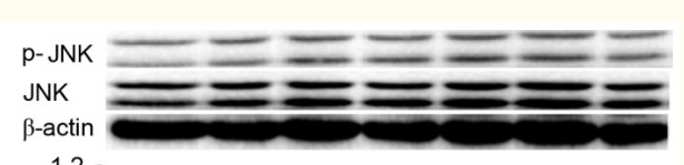
- Western Blot analysis of various cells using Phospho-JNK1/2/3 (T183/Y185) Polyclonal Antibody diluted at 1:2000
.jpg)
- Western Blot analysis of 293 cells using Phospho-JNK1/2/3 (T183/Y185) Polyclonal Antibody diluted at 1:2000
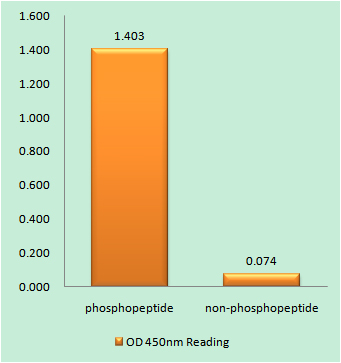
- Enzyme-Linked Immunosorbent Assay (Phospho-ELISA) for Immunogen Phosphopeptide (Phospho-left) and Non-Phosphopeptide (Phospho-right), using JNK1/2/3 (Phospho-Thr183+Tyr185) Antibody
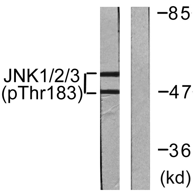
- Western blot analysis of lysates from 293 cells treated with UV 5', using JNK1/2/3 (Phospho-Thr183+Tyr185) Antibody. The lane on the right is blocked with the phospho peptide.


