CHOP mouse Monoclonal Antibody(2B1)
- Catalog No.:YM3668
- Applications:WB;IF;IHC
- Reactivity:Human;Rat;Mouse
- Target:
- CHOP
- Fields:
- >>MAPK signaling pathway;>>Protein processing in endoplasmic reticulum;>>Apoptosis;>>Non-alcoholic fatty liver disease;>>Alzheimer disease;>>Parkinson disease;>>Amyotrophic lateral sclerosis;>>Prion disease;>>Pathways of neurodegeneration - multiple diseases;>>Transcriptional misregulation in cancer;>>Lipid and atherosclerosis
- Gene Name:
- DDIT3
- Protein Name:
- DDIT3
- Human Gene Id:
- 1649
- Human Swiss Prot No:
- P35638
- Mouse Swiss Prot No:
- P35639
- Rat Swiss Prot No:
- Q62857
- Immunogen:
- Synthetic Peptide of CHOP at AA range of 10-90
- Specificity:
- CHOP protein detects endogenous levels of DDIT3
- Formulation:
- Liquid in PBS containing 50% glycerol, 0.5% BSA and 0.02% sodium azide.
- Source:
- Monoclonal, Mouse
- Dilution:
- WB 1:1000-2000, IHC 1:100-200 IF 1:200
- Purification:
- The antibody was affinity-purified from mouse ascites by affinity-chromatography using specific immunogen.
- Concentration:
- 1 mg/ml
- Storage Stability:
- -15°C to -25°C/1 year(Do not lower than -25°C)
- Other Name:
- DDIT3
- Observed Band(KD):
- 27kD
- Background:
- This gene encodes a member of the CCAAT/enhancer-binding protein (C/EBP) family of transcription factors. The protein functions as a dominant-negative inhibitor by forming heterodimers with other C/EBP members, such as C/EBP and LAP (liver activator protein), and preventing their DNA binding activity. The protein is implicated in adipogenesis and erythropoiesis, is activated by endoplasmic reticulum stress, and promotes apoptosis. Fusion of this gene and FUS on chromosome 16 or EWSR1 on chromosome 22 induced by translocation generates chimeric proteins in myxoid liposarcomas or Ewing sarcoma. Multiple alternatively spliced transcript variants encoding two isoforms with different length have been identified. [provided by RefSeq, Aug 2010],
- Function:
- disease:A chromosomal aberration involving DDIT3 is found in a form of malignant myxoid liposarcoma [MIM:126337]. Translocation t(12;16)(q13;p11) with FUS.,function:Inhibits the DNA-binding activity of C/EBP and LAP by forming heterodimers that cannot bind DNA.,similarity:Belongs to the bZIP family.,similarity:Contains 1 bZIP domain.,subunit:Heterodimer.,
- Subcellular Location:
- Cytoplasm . Nucleus . Present in the cytoplasm under non-stressed conditions and ER stress leads to its nuclear accumulation. .
- Expression:
- Muscle,Skeletal muscle,
Activation of the PERK-CHOP signaling pathway during endoplasmic reticulum stress contributes to olanzapine-induced dyslipidemia. ACTA PHARMACOLOGICA SINICA Chang-hua Hu WB Mouse,Human liver HepG2 cell
- June 19-2018
- WESTERN IMMUNOBLOTTING PROTOCOL
- June 19-2018
- IMMUNOHISTOCHEMISTRY-PARAFFIN PROTOCOL
- June 19-2018
- IMMUNOFLUORESCENCE PROTOCOL
- September 08-2020
- FLOW-CYTOMEYRT-PROTOCOL
- May 20-2022
- Cell-Based ELISA│解您多样本WB检测之困扰
- July 13-2018
- CELL-BASED-ELISA-PROTOCOL-FOR-ACETYL-PROTEIN
- July 13-2018
- CELL-BASED-ELISA-PROTOCOL-FOR-PHOSPHO-PROTEIN
- July 13-2018
- Antibody-FAQs
- Products Images
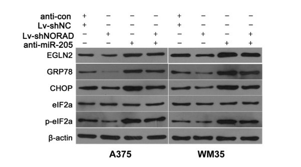
- Chen, Yong, et al. "Overexpression of long non‐coding RNA NORAD promotes invasion and migration in malignant melanoma via regulating the MIR‐205‐EGLN2 pathway." Cancer medicine (2019).

- Immunofluorescence analysis of Hela cell. 1,Calnexin Polyclonal Antibody(green) was diluted at 1:200(4° overnight). (red) was diluted at 1:200(4° overnight). 2, Goat Anti Rabbit Alexa Fluor 488 Catalog:RS3211 was diluted at 1:1000(room temperature, 50min). Goat Anti Mouse Alexa Fluor 594 Catalog:RS3608 was diluted at 1:1000(room temperature, 50min).
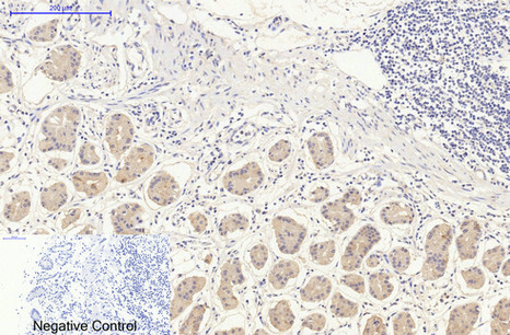
- Immunohistochemical analysis of paraffin-embedded Human-stomach tissue. 1,CHOP Mouse Monoclonal Antibody(2B1) was diluted at 1:200(4°C,overnight). 2, Sodium citrate pH 6.0 was used for antibody retrieval(>98°C,20min). 3,Secondary antibody was diluted at 1:200(room tempeRature, 30min). Negative control was used by secondary antibody only.
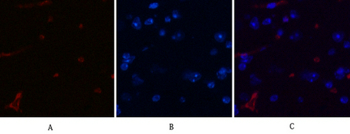
- Immunofluorescence analysis of Mouse-brain tissue. 1,CHOP Mouse Monoclonal Antibody(2B1)(red) was diluted at 1:200(4°C,overnight). 2, Cy3 labled Secondary antibody was diluted at 1:300(room temperature, 50min).3, Picture B: DAPI(blue) 10min. Picture A:Target. Picture B: DAPI. Picture C: merge of A+B
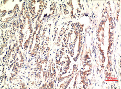
- Immunohistochemical analysis of paraffin-embedded Human Stomach Carcinoma Tissue using CHOP Mouse mAb diluted at 1:200.
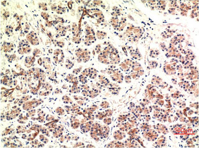
- Immunohistochemical analysis of paraffin-embedded Human Pancreas Carcinoma Tissue using CHOP Mouse mAb diluted at 1:200.
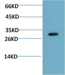
- Western blot analysis of Mouse Liver Tissue Lysate using CHOP Mouse mAb diluted at 1:2000.


