CD2 Monoclonal Antibody(3F3)
- Catalog No.:YM3069
- Applications:IHC;IF
- Reactivity:Human;Mouse;Rat
- Target:
- CD2
- Fields:
- >>Cell adhesion molecules;>>Hematopoietic cell lineage
- Gene Name:
- CD2
- Protein Name:
- T-cell surface antigen CD2
- Human Gene Id:
- 914
- Human Swiss Prot No:
- P06729
- Mouse Gene Id:
- 12481
- Mouse Swiss Prot No:
- P08920
- Rat Gene Id:
- 497761
- Rat Swiss Prot No:
- P08921
- Immunogen:
- Synthetic Peptide of CD2
- Specificity:
- The antibody detects endogenous CD2 proteins.
- Formulation:
- PBS, pH 7.4, containing 0.5%BSA, 0.02% sodium azide as Preservative and 50% Glycerol.
- Source:
- Monoclonal, Mouse
- Dilution:
- IHC 1:200. IF 1:50-200
- Purification:
- The antibody was affinity-purified from mouse ascites by affinity-chromatography using specific immunogen.
- Storage Stability:
- -15°C to -25°C/1 year(Do not lower than -25°C)
- Other Name:
- CD2;SRBC;T-cell surface antigen CD2;Erythrocyte receptor;LFA-2;LFA-3 receptor;Rosette receptor;T-cell surface antigen T11/Leu-5;CD2
- Molecular Weight(Da):
- 39kD
- Background:
- The protein encoded by this gene is a surface antigen found on all peripheral blood T-cells. The encoded protein interacts with LFA3 (CD58) on antigen presenting cells to optimize immune recognition. A locus control region (LCR) has been found in the 3' flanking sequence of this gene. [provided by RefSeq, Jun 2016],
- Function:
- function:CD2 interacts with lymphocyte function-associated antigen (LFA-3) and CD48/BCM1 to mediate adhesion between T-cells and other cell types. CD2 is implicated in the triggering of T-cells, the cytoplasmic domain is implicated in the signaling function.,online information:CD2 entry,similarity:Contains 1 Ig-like C2-type (immunoglobulin-like) domain.,similarity:Contains 1 Ig-like V-type (immunoglobulin-like) domain.,subunit:Interacts with CD2AP (By similarity). Interacts with PSTPIP1.,
- Subcellular Location:
- Cell membrane ; Single-pass type I membrane protein .
- Expression:
- Expressed in natural killer cells (at protein level).
- June 19-2018
- WESTERN IMMUNOBLOTTING PROTOCOL
- June 19-2018
- IMMUNOHISTOCHEMISTRY-PARAFFIN PROTOCOL
- June 19-2018
- IMMUNOFLUORESCENCE PROTOCOL
- September 08-2020
- FLOW-CYTOMEYRT-PROTOCOL
- May 20-2022
- Cell-Based ELISA│解您多样本WB检测之困扰
- July 13-2018
- CELL-BASED-ELISA-PROTOCOL-FOR-ACETYL-PROTEIN
- July 13-2018
- CELL-BASED-ELISA-PROTOCOL-FOR-PHOSPHO-PROTEIN
- July 13-2018
- Antibody-FAQs
- Products Images

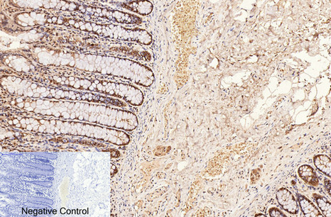
- Immunohistochemical analysis of paraffin-embedded Human-colon tissue. 1,CD2 Monoclonal Antibody(3F3) was diluted at 1:200(4°C,overnight). 2, Sodium citrate pH 6.0 was used for antibody retrieval(>98°C,20min). 3,Secondary antibody was diluted at 1:200(room tempeRature, 30min). Negative control was used by secondary antibody only.

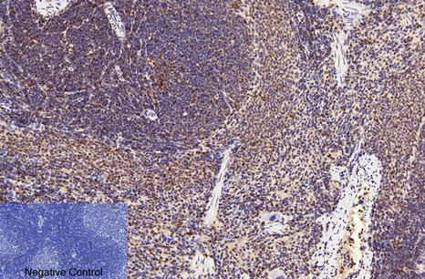
- Immunohistochemical analysis of paraffin-embedded Rat-spleen tissue. 1,CD2 Monoclonal Antibody(3F3) was diluted at 1:200(4°C,overnight). 2, Sodium citrate pH 6.0 was used for antibody retrieval(>98°C,20min). 3,Secondary antibody was diluted at 1:200(room tempeRature, 30min). Negative control was used by secondary antibody only.

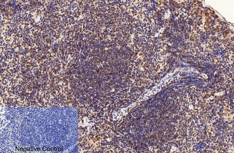
- Immunohistochemical analysis of paraffin-embedded Mouse-spleen tissue. 1,CD2 Monoclonal Antibody(3F3) was diluted at 1:200(4°C,overnight). 2, Sodium citrate pH 6.0 was used for antibody retrieval(>98°C,20min). 3,Secondary antibody was diluted at 1:200(room tempeRature, 30min). Negative control was used by secondary antibody only.

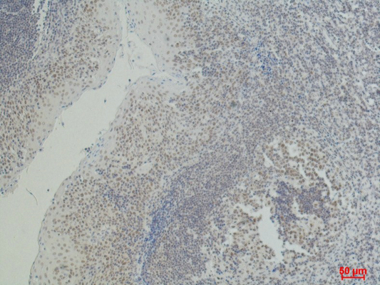
- Immunohistochemical analysis of paraffin-embedded Human Tonsil Caricnoma using CD2 Mouse mAb diluted at 1:500.


