Arg Monoclonal Antibody
- Catalog No.:YM0046
- Applications:WB;IF;ELISA
- Reactivity:Human;Mouse
- Target:
- Arg
- Fields:
- >>ErbB signaling pathway;>>Ras signaling pathway;>>Chemical carcinogenesis - reactive oxygen species;>>Viral myocarditis
- Gene Name:
- ABL2
- Protein Name:
- Tyrosine-protein kinase ABL2
- Human Gene Id:
- 27
- Human Swiss Prot No:
- P42684
- Mouse Swiss Prot No:
- Q4JIM5
- Immunogen:
- Purified recombinant fragment of Arg expressed in E. Coli.
- Specificity:
- Arg Monoclonal Antibody detects endogenous levels of Arg protein.
- Formulation:
- Liquid in PBS containing 50% glycerol, 0.5% BSA and 0.02% sodium azide.
- Source:
- Monoclonal, Mouse
- Dilution:
- WB 1:500 - 1:2000. IF 1:200 - 1:1000. ELISA: 1:10000. Not yet tested in other applications.
- Purification:
- Affinity purification
- Storage Stability:
- -15°C to -25°C/1 year(Do not lower than -25°C)
- Other Name:
- ABL2;ABLL;ARG;Abelson tyrosine-protein kinase 2;Abelson murine leukemia viral oncogene homolog 2;Abelson-related gene protein;Tyrosine-protein kinase ARG
- Molecular Weight(Da):
- 128kD
- References:
- 1. Yoshimi I, Takashi I, Tsuneyuki O, et al. Blood. 2000; 95(6): 2126-2131.
2. Scheijen, B. and Griffin, J.D. Oncogene. 2002); 21:3314-33.
- Background:
- This gene encodes a member of the Abelson family of nonreceptor tyrosine protein kinases. The protein is highly similar to the c-abl oncogene 1 protein, including the tyrosine kinase, SH2 and SH3 domains, and it plays a role in cytoskeletal rearrangements through its C-terminal F-actin- and microtubule-binding sequences. This gene is expressed in both normal and tumor cells, and is involved in translocation with the ets variant 6 gene in leukemia. Multiple alternatively spliced transcript variants encoding different protein isoforms have been found for this gene. [provided by RefSeq, Nov 2009],
- Function:
- catalytic activity:ATP + a [protein]-L-tyrosine = ADP + a [protein]-L-tyrosine phosphate.,caution:The sequence shown here is derived from an Ensembl automatic analysis pipeline and should be considered as preliminary data.,cofactor:Magnesium or manganese.,enzyme regulation:Stabilized in the inactive form by an association between the SH3 domain and the SH2-TK linker region, interactions of the amino-terminal cap, and contributions from an amino-terminal myristoyl group and phospholipids. Activated by autophosphorylation as well as by SRC-family kinase-mediated phosphorylation. Activated by RIN1 binding to the SH2 and SH3 domains. Inhibited by imatinib mesylate (Gleevec) which is used for the treatment of chronic myeloid leukemia (CML).,function:Regulates cytoskeleton remodeling during cell differentiation, cell division and cell adhesion. Localizes to dynamic actin structures, and phosph
- Subcellular Location:
- Cytoplasm, cytoskeleton.
- Expression:
- Widely expressed.
- June 19-2018
- WESTERN IMMUNOBLOTTING PROTOCOL
- June 19-2018
- IMMUNOHISTOCHEMISTRY-PARAFFIN PROTOCOL
- June 19-2018
- IMMUNOFLUORESCENCE PROTOCOL
- September 08-2020
- FLOW-CYTOMEYRT-PROTOCOL
- May 20-2022
- Cell-Based ELISA│解您多样本WB检测之困扰
- July 13-2018
- CELL-BASED-ELISA-PROTOCOL-FOR-ACETYL-PROTEIN
- July 13-2018
- CELL-BASED-ELISA-PROTOCOL-FOR-PHOSPHO-PROTEIN
- July 13-2018
- Antibody-FAQs
- Products Images

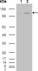
- Western Blot analysis using Arg Monoclonal Antibody against HEK293T cells transfected with the pCMV6-ENTRY control (1) and pCMV6-ENTRY ABL2 cDNA (2).

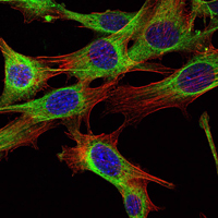
- Immunofluorescence analysis of NIH/3T3 cells using Arg Monoclonal Antibody (green). Blue: DRAQ5 fluorescent DNA dye. Red: Actin filaments have been labeled with Alexa Fluor-555 phalloidin.


