GDF11 rabbit pAb
- Catalog No.:YT6928
- Applications:WB
- Reactivity:Human;Mouse;Rat
- Target:
- GDF11
- Fields:
- >>Cytokine-cytokine receptor interaction
- Gene Name:
- GDF11 BMP11
- Protein Name:
- GDF11
- Human Gene Id:
- 10220
- Human Swiss Prot No:
- O95390
- Mouse Gene Id:
- 14561
- Mouse Swiss Prot No:
- Q9Z1W4
- Rat Swiss Prot No:
- Q9Z217
- Immunogen:
- Synthesized peptide derived from human GDF11 AA range: 217-267
- Specificity:
- This antibody detects endogenous levels of GDF11 at Human/Mouse/Rat
- Formulation:
- Liquid in PBS containing 50% glycerol, 0.5% BSA and 0.02% sodium azide.
- Source:
- Polyclonal, Rabbit,IgG
- Dilution:
- WB 1:500-2000
- Purification:
- The antibody was affinity-purified from rabbit antiserum by affinity-chromatography using epitope-specific immunogen.
- Concentration:
- 1 mg/ml
- Storage Stability:
- -15°C to -25°C/1 year(Do not lower than -25°C)
- Molecular Weight(Da):
- 45kD
- Background:
- This gene encodes a secreted ligand of the TGF-beta (transforming growth factor-beta) superfamily of proteins. Ligands of this family bind various TGF-beta receptors leading to recruitment and activation of SMAD family transcription factors that regulate gene expression. The encoded preproprotein is proteolytically processed to generate each subunit of the disulfide-linked homodimer. This protein plays a role in the development of the nervous and other organ systems, and may regulate aging. [provided by RefSeq, Aug 2016],
- Function:
- function:Secreted signal that acts globally to specify positional identity along the anterior/posterior axis during development. Play critical roles in patterning both mesodermal and neural tissues and in establishing the skeletal pattern.,similarity:Belongs to the TGF-beta family.,subunit:Homodimer; disulfide-linked.,
- Subcellular Location:
- Secreted .
- Expression:
- In the embryo, strong expression is seen in the palatal epithelia, including the medial edge epithelial and midline epithelial seam of the palatal shelves. Less pronounced expression is also seen throughout the palatal shelf and tongue mesenchyme.
- June 19-2018
- WESTERN IMMUNOBLOTTING PROTOCOL
- June 19-2018
- IMMUNOHISTOCHEMISTRY-PARAFFIN PROTOCOL
- June 19-2018
- IMMUNOFLUORESCENCE PROTOCOL
- September 08-2020
- FLOW-CYTOMEYRT-PROTOCOL
- May 20-2022
- Cell-Based ELISA│解您多样本WB检测之困扰
- July 13-2018
- CELL-BASED-ELISA-PROTOCOL-FOR-ACETYL-PROTEIN
- July 13-2018
- CELL-BASED-ELISA-PROTOCOL-FOR-PHOSPHO-PROTEIN
- July 13-2018
- Antibody-FAQs
- Products Images
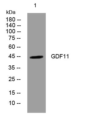
- Western blot analysis of lysates from A549 cells, primary antibody was diluted at 1:1000, 4°over night


