- Home
- About
- Promotions
-
Products
-
Elisa Kits
- |
-
Primary antibodies
- |
-
Secondary antibodies
- |
-
Proteins
- |
-
IHC reagents
- |
-
WB reagents
- PonceauS Staining Solution
- PBST Washing Buffer, 10X
- 1.5M Tris-HCl Buffer, pH8.8
- 1M Tris-HCl Buffer, pH6.8
- 10% SDS Solution
- Prestained Protein Marker
- TBST Washing Buffer, 10X
- SDS PAGE Loading Buffer, 5X
- Stripping Buffered Solution
- Tris Buffer, pH7.4, 10X
- Total Protein Extraction Kit
- Running Buffer, 10X
- Transfer Buffer, 10X
- 30% Acr-Bis(29:1) Solution
- Tris电泳液速溶颗粒
- PBS(1X, premixed powder)
- TBS(1X, premixed powder)
- 快速封闭液
- 转膜液速溶颗粒
- Chemical reagents
- News
- Distributor
- Resources
- Contact
- Home
- >
- Info
- >
- CETN1 rabbit pAb
- >
- Go Back
CETN1 rabbit pAb
- Catalog No.:YT6644
- Applications:WB
- Reactivity:Human;Mouse
- Gene Name:
- CETN1 CEN1 CETN
- Immunogen:
- Synthesized peptide derived from human CETN1 AA range: 96-146
- Specificity:
- This antibody detects endogenous levels of CETN1 at Human/Mouse
- Formulation:
- Liquid in PBS containing 50% glycerol, 0.5% BSA and 0.02% sodium azide.
- Source:
- Polyclonal, Rabbit,IgG
- Purification:
- The antibody was affinity-purified from rabbit antiserum by affinity-chromatography using epitope-specific immunogen.
- Storage Stability:
- -15°C to -25°C/1 year(Do not lower than -25°C)
- Molecular Weight(Da):
- 19kD
- Background:
- The protein encoded by this gene plays important roles in the determination of centrosome position and segregation, and in the process of microtubule severing. This protein is localized to the centrosome of interphase cells, and redistributes to the region of the spindle poles during mitosis, reflecting the dynamic behavior of the centrosome during the cell cycle. [provided by RefSeq, Jan 2015],
- Function:
- function:Plays a fundamental role in microtubule-organizing center structure and function.,miscellaneous:Binds two moles of calcium per mole of protein.,similarity:Belongs to the centrin family.,similarity:Contains 4 EF-hand domains.,subcellular location:Centrosome of interphase and mitotic cells.,subunit:Monomer.,
- Subcellular Location:
- Cytoplasm, cytoskeleton, microtubule organizing center, centrosome . Cell projection, cilium . Centrosome of interphase and mitotic cells. In the retinal photoreceptor cells, localizes at the connecting cilium, a thin bridge linking the cell body and the light-sensing outer segment (By similarity). .
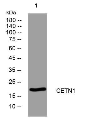
- Western blot analysis of lysates from K562 cells, primary antibody was diluted at 1:1000, 4°over night


