LCOR rabbit pAb
- Catalog No.:YT6380
- Applications:WB;IHC
- Reactivity:Human;Mouse
- Target:
- LCOR
- Gene Name:
- LCOR KIAA1795 MLR2
- Protein Name:
- LCOR
- Human Gene Id:
- 84458
- Human Swiss Prot No:
- Q96JN0
- Mouse Gene Id:
- 212391
- Mouse Swiss Prot No:
- Q6ZPI3
- Immunogen:
- Synthesized peptide derived from human LCOR AA range: 244-294
- Specificity:
- This antibody detects endogenous levels of LCOR at Human/Mouse
- Formulation:
- Liquid in PBS containing 50% glycerol, 0.5% BSA and 0.02% sodium azide.
- Source:
- Polyclonal, Rabbit,IgG
- Dilution:
- WB 1:500-2000;IHC 1:50-300
- Purification:
- The antibody was affinity-purified from rabbit antiserum by affinity-chromatography using epitope-specific immunogen.
- Concentration:
- 1 mg/ml
- Storage Stability:
- -15°C to -25°C/1 year(Do not lower than -25°C)
- Molecular Weight(Da):
- 48kD
- Background:
- LCOR is a transcriptional corepressor widely expressed in fetal and adult tissues that is recruited to agonist-bound nuclear receptors through a single LxxLL motif, also referred to as a nuclear receptor (NR) box (Fernandes et al., 2003 [PubMed 12535528]).[supplied by OMIM, Mar 2008],
- Function:
- function:May act as transcription activator that binds DNA elements with the sequence 5'-CCCTATCGATCGATCTCTACCT-3' (By similarity). Repressor of ligand-dependent transcription activation by target nuclear receptors. Repressor of ligand-dependent transcription activation by ESR1, ESR2, NR3C1, PGR, RARA, RARB, RARG, RXRA and VDR.,similarity:Contains 1 HTH psq-type DNA-binding domain.,subunit:Interacts with ESR1 and ESR2 in the presence of estradiol. Interacts with CTBP1, HDAC3 and HDAC6. Component of a large corepressor complex that contains about 20 proteins, including CTBP1, CTBP2, HDAC1 and HDAC2.,tissue specificity:Ubiquitous.,
- Subcellular Location:
- Nucleus .
- Expression:
- Ubiquitous.
- June 19-2018
- WESTERN IMMUNOBLOTTING PROTOCOL
- June 19-2018
- IMMUNOHISTOCHEMISTRY-PARAFFIN PROTOCOL
- June 19-2018
- IMMUNOFLUORESCENCE PROTOCOL
- September 08-2020
- FLOW-CYTOMEYRT-PROTOCOL
- May 20-2022
- Cell-Based ELISA│解您多样本WB检测之困扰
- July 13-2018
- CELL-BASED-ELISA-PROTOCOL-FOR-ACETYL-PROTEIN
- July 13-2018
- CELL-BASED-ELISA-PROTOCOL-FOR-PHOSPHO-PROTEIN
- July 13-2018
- Antibody-FAQs
- Products Images
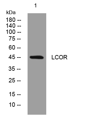
- Western blot analysis of lysates from HpeG2 cells, primary antibody was diluted at 1:1000, 4°over night
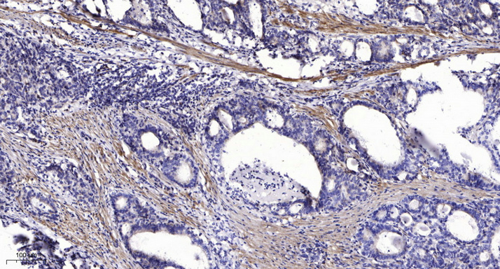
- Immunohistochemical analysis of paraffin-embedded human Gastric adenocarcinoma. 1, Antibody was diluted at 1:200(4° overnight). 2, Tris-EDTA,pH9.0 was used for antigen retrieval. 3,Secondary antibody was diluted at 1:200(room temperature, 45min).


