GDPD1 rabbit pAb
- Catalog No.:YT7770
- Applications:WB
- Reactivity:Human;Mouse;Rat
- Target:
- GDPD1
- Fields:
- >>Ether lipid metabolism;>>Metabolic pathways
- Gene Name:
- GDPD1 GDE4
- Protein Name:
- GDPD1
- Human Gene Id:
- 284161
- Human Swiss Prot No:
- Q8N9F7
- Mouse Gene Id:
- 66569
- Mouse Swiss Prot No:
- Q9CRY7
- Rat Gene Id:
- 303407
- Rat Swiss Prot No:
- Q0VGK4
- Immunogen:
- Synthesized peptide derived from human GDPD1 AA range: 247-297
- Specificity:
- This antibody detects endogenous levels of GDPD1 at Human/Mouse/Rat
- Formulation:
- Liquid in PBS containing 50% glycerol, 0.5% BSA and 0.02% sodium azide.
- Source:
- Polyclonal, Rabbit,IgG
- Dilution:
- WB 1:500-2000
- Purification:
- The antibody was affinity-purified from rabbit antiserum by affinity-chromatography using epitope-specific immunogen.
- Concentration:
- 1 mg/ml
- Storage Stability:
- -15°C to -25°C/1 year(Do not lower than -25°C)
- Molecular Weight(Da):
- 35kD
- Background:
- This gene encodes a member of the glycerophosphodiester phosphodiesterase family of enzymes that catalyze the hydrolysis of deacylated glycerophospholipids to glycerol phosphate and alcohol. The encoded protein is localized to the cytoplasm and concentrates near the perinuclear region. Alternative splicing results in multiple transcript variants. [provided by RefSeq, Oct 2009],
- Function:
- similarity:Belongs to the glycerophosphoryl diester phosphodiesterase family.,similarity:Contains 1 GDPD domain.,tissue specificity:Detected in placenta, liver, kidney, pancreas, spleen, thymus, ovary, small intestine and peripheral blood leukocytes.,
- Subcellular Location:
- Cytoplasm . Membrane ; Multi-pass membrane protein . Cytoplasm, perinuclear region . Endoplasmic reticulum . Concentrated at the perinuclear region and the cell periphery (PubMed:18991142). .
- Expression:
- Widely expressed with high expression level in testis.
- June 19-2018
- WESTERN IMMUNOBLOTTING PROTOCOL
- June 19-2018
- IMMUNOHISTOCHEMISTRY-PARAFFIN PROTOCOL
- June 19-2018
- IMMUNOFLUORESCENCE PROTOCOL
- September 08-2020
- FLOW-CYTOMEYRT-PROTOCOL
- May 20-2022
- Cell-Based ELISA│解您多样本WB检测之困扰
- July 13-2018
- CELL-BASED-ELISA-PROTOCOL-FOR-ACETYL-PROTEIN
- July 13-2018
- CELL-BASED-ELISA-PROTOCOL-FOR-PHOSPHO-PROTEIN
- July 13-2018
- Antibody-FAQs
- Products Images
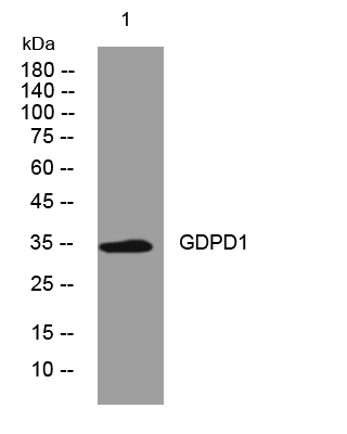
- Western blot analysis of lysates from VEC cells, primary antibody was diluted at 1:1000, 4°over night


