LEO1 rabbit pAb
- Catalog No.:YT7689
- Applications:WB;ELISA;IHC
- Reactivity:Human;Mouse;Rat
- Target:
- LEO1
- Gene Name:
- LEO1 RDL
- Protein Name:
- LEO1
- Human Gene Id:
- 123169
- Human Swiss Prot No:
- Q8WVC0
- Mouse Gene Id:
- 235497
- Mouse Swiss Prot No:
- Q5XJE5
- Rat Gene Id:
- 300837
- Rat Swiss Prot No:
- Q641X2
- Immunogen:
- Synthesized peptide derived from human LEO1 AA range: 159-209
- Specificity:
- This antibody detects endogenous levels of LEO1 at Human/Mouse/Rat
- Formulation:
- Liquid in PBS containing 50% glycerol, 0.5% BSA and 0.02% sodium azide.
- Source:
- Polyclonal, Rabbit,IgG
- Dilution:
- WB 1:500-2000;IHC 1:50-300; ELISA 2000-20000
- Purification:
- The antibody was affinity-purified from rabbit antiserum by affinity-chromatography using epitope-specific immunogen.
- Concentration:
- 1 mg/ml
- Storage Stability:
- -15°C to -25°C/1 year(Do not lower than -25°C)
- Molecular Weight(Da):
- 73kD
- Background:
- LEO1, parafibromin (CDC73; MIM 607393), CTR9 (MIM 609366), and PAF1 (MIM 610506) form the PAF protein complex that associates with the RNA polymerase II subunit POLR2A (MIM 180660) and with a histone methyltransferase complex (Rozenblatt-Rosen et al., 2005 [PubMed 15632063]).[supplied by OMIM, Mar 2008],
- Function:
- function:The PAF1 complex is a multifunctional complex. The PAF1 complex interacts with POLR2A. May be involved in both initiation and elongation, histone methylation and RNA processing. Overexpression of LEO1 induces cell growth arrest and premature senescence of fibroblasts.,similarity:Belongs to the LEO1 family.,subunit:Component of the PAF1 complex, which consists of at least of CDC73, PAF1, LEO1 and CTR9. Interacts with CDC73.,tissue specificity:Highly expressed in skeletal muscle and heart. Weakly expressed in placenta and liver.,
- Subcellular Location:
- Nucleus .
- Expression:
- Highly expressed in skeletal muscle and heart. Weakly expressed in placenta and liver.
- June 19-2018
- WESTERN IMMUNOBLOTTING PROTOCOL
- June 19-2018
- IMMUNOHISTOCHEMISTRY-PARAFFIN PROTOCOL
- June 19-2018
- IMMUNOFLUORESCENCE PROTOCOL
- September 08-2020
- FLOW-CYTOMEYRT-PROTOCOL
- May 20-2022
- Cell-Based ELISA│解您多样本WB检测之困扰
- July 13-2018
- CELL-BASED-ELISA-PROTOCOL-FOR-ACETYL-PROTEIN
- July 13-2018
- CELL-BASED-ELISA-PROTOCOL-FOR-PHOSPHO-PROTEIN
- July 13-2018
- Antibody-FAQs
- Products Images
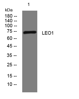
- Western blot analysis of lysates from U2OS cells, primary antibody was diluted at 1:1000, 4°over night
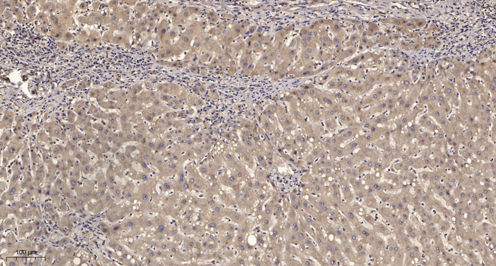
- Immunohistochemical analysis of paraffin-embedded human liver cancer. 1, Antibody was diluted at 1:200(4° overnight). 2, Tris-EDTA,pH9.0 was used for antigen retrieval. 3,Secondary antibody was diluted at 1:200(room temperature, 45min).


