DREB rabbit pAb
- Catalog No.:YT7666
- Applications:WB
- Reactivity:Human;Mouse;Rat
- Target:
- DREB
- Gene Name:
- DBN1 D0S117E
- Protein Name:
- DREB
- Human Gene Id:
- 1627
- Human Swiss Prot No:
- Q16643
- Mouse Gene Id:
- 56320
- Mouse Swiss Prot No:
- Q9QXS6
- Rat Gene Id:
- 81653
- Rat Swiss Prot No:
- Q07266
- Immunogen:
- Synthesized peptide derived from human DREB AA range: 484-534
- Specificity:
- This antibody detects endogenous levels of DREB at Human/Mouse/Rat
- Formulation:
- Liquid in PBS containing 50% glycerol, 0.5% BSA and 0.02% sodium azide.
- Source:
- Polyclonal, Rabbit,IgG
- Dilution:
- WB 1:500-2000
- Purification:
- The antibody was affinity-purified from rabbit antiserum by affinity-chromatography using epitope-specific immunogen.
- Concentration:
- 1 mg/ml
- Storage Stability:
- -15°C to -25°C/1 year(Do not lower than -25°C)
- Molecular Weight(Da):
- 71kD
- Background:
- The protein encoded by this gene is a cytoplasmic actin-binding protein thought to play a role in the process of neuronal growth. It is a member of the drebrin family of proteins that are developmentally regulated in the brain. A decrease in the amount of this protein in the brain has been implicated as a possible contributing factor in the pathogenesis of memory disturbance in Alzheimer's disease. At least two alternative splice variants encoding different protein isoforms have been described for this gene. [provided by RefSeq, Jul 2008],
- Function:
- function:Drebrins might play some role in cell migration, extension of neuronal processes and plasticity of dendrites, respectively.,similarity:Contains 1 ADF-H domain.,subunit:Binds F-actin.,tissue specificity:Brain neurons. Also found in the heart, placenta, skeletal muscle, kidney and pancreas.,
- Subcellular Location:
- Cytoplasm . Cell projection, dendrite . Cytoplasm, cell cortex . Cell junction . Cell projection, growth cone . In the absence of antigen, evenly distributed throughout subcortical regions of the T-cell membrane and cytoplasm (PubMed:20215400). In the presence of antigen, distributes to the immunological synapse forming at the T-cell-APC contact area, where it localizes at the peripheral and distal supramolecular activation clusters (SMAC) (PubMed:20215400). Colocalized with RUFY3 and F-actin at the transitional domain of the axonal growth cone (By similarity). .
- Expression:
- Expressed in the brain, with expression in the molecular layer of the dentate gyrus, stratum pyramidale, and stratum radiatum of the hippocampus (at protein level) (PubMed:8838578). Also expressed in the terminal varicosities distributed along dendritic trees of pyramidal cells in CA4 and CA3 of the hippocampus (at protein level) (PubMed:8838578). Expressed in pyramidal cells in CA2, CA1 and the subiculum of the hippocampus (at protein level) (PubMed:8838578). Expressed in peripheral blood lymphocytes, including T-cells (at protein level) (PubMed:20215400). Expressed in the brain (PubMed:8216329, Ref.2). Expressed in the heart, placenta, lung, skeletal muscle, kidney, pancreas, skin fibroblasts, gingival fibroblasts and bone-derived cells (Ref.2).
- June 19-2018
- WESTERN IMMUNOBLOTTING PROTOCOL
- June 19-2018
- IMMUNOHISTOCHEMISTRY-PARAFFIN PROTOCOL
- June 19-2018
- IMMUNOFLUORESCENCE PROTOCOL
- September 08-2020
- FLOW-CYTOMEYRT-PROTOCOL
- May 20-2022
- Cell-Based ELISA│解您多样本WB检测之困扰
- July 13-2018
- CELL-BASED-ELISA-PROTOCOL-FOR-ACETYL-PROTEIN
- July 13-2018
- CELL-BASED-ELISA-PROTOCOL-FOR-PHOSPHO-PROTEIN
- July 13-2018
- Antibody-FAQs
- Products Images
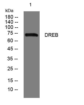
- Western blot analysis of lysates from Hela cells, primary antibody was diluted at 1:1000, 4°over night


