TRI11 rabbit pAb
- Catalog No.:YT7631
- Applications:WB
- Reactivity:Human;Mouse;Rat
- Target:
- TRI11
- Gene Name:
- TRIM11 RNF92
- Protein Name:
- TRI11
- Human Gene Id:
- 81559
- Human Swiss Prot No:
- Q96F44
- Mouse Gene Id:
- 338364
- Mouse Swiss Prot No:
- Q99PQ2
- Rat Swiss Prot No:
- B1H278
- Immunogen:
- Synthesized peptide derived from human TRI11 AA range: 128-178
- Specificity:
- This antibody detects endogenous levels of TRI11 at Human/Mouse/Rat
- Formulation:
- Liquid in PBS containing 50% glycerol, 0.5% BSA and 0.02% sodium azide.
- Source:
- Polyclonal, Rabbit,IgG
- Dilution:
- WB 1:500-2000
- Purification:
- The antibody was affinity-purified from rabbit antiserum by affinity-chromatography using epitope-specific immunogen.
- Concentration:
- 1 mg/ml
- Storage Stability:
- -15°C to -25°C/1 year(Do not lower than -25°C)
- Molecular Weight(Da):
- 51kD
- Background:
- The protein encoded by this gene is a member of the tripartite motif (TRIM) family. The TRIM motif includes three zinc-binding domains, a RING, a B-box type 1 and a B-box type 2, and a coiled-coil region. This protein localizes to the nucleus and the cytoplasm. Its function has not been identified. [provided by RefSeq, Jul 2008],
- Function:
- domain:The coiled-coil domain and the B30.2 domain are both necessary for interaction with humanin.,function:May have E3 ubiquitin ligase activity. May contribute to the regulation of the intracellular level of humanin or humanin-containing proteins through the proteasomal degradation pathway.,similarity:Belongs to the TRIM/RBCC family.,similarity:Contains 1 B box-type zinc finger.,similarity:Contains 1 B30.2/SPRY domain.,similarity:Contains 1 RING-type zinc finger.,subunit:Binds cytoplasmic tail of integrin alpha-1. Interacts with the humanin peptide.,tissue specificity:Ubiquitous.,
- Subcellular Location:
- Cytoplasm. Nucleus.
- Expression:
- Ubiquitous.
- June 19-2018
- WESTERN IMMUNOBLOTTING PROTOCOL
- June 19-2018
- IMMUNOHISTOCHEMISTRY-PARAFFIN PROTOCOL
- June 19-2018
- IMMUNOFLUORESCENCE PROTOCOL
- September 08-2020
- FLOW-CYTOMEYRT-PROTOCOL
- May 20-2022
- Cell-Based ELISA│解您多样本WB检测之困扰
- July 13-2018
- CELL-BASED-ELISA-PROTOCOL-FOR-ACETYL-PROTEIN
- July 13-2018
- CELL-BASED-ELISA-PROTOCOL-FOR-PHOSPHO-PROTEIN
- July 13-2018
- Antibody-FAQs
- Products Images
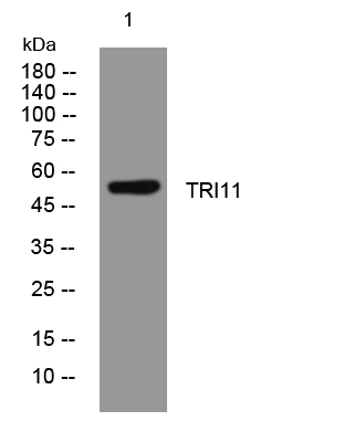
- Western blot analysis of lysates from HpeG2 cells, primary antibody was diluted at 1:1000, 4°over night


