ELMO3 rabbit pAb
- Catalog No.:YT7574
- Applications:WB
- Reactivity:Human;Mouse;Rat
- Target:
- ELMO3
- Fields:
- >>Bacterial invasion of epithelial cells
- Gene Name:
- ELMO3
- Protein Name:
- ELMO3
- Human Gene Id:
- 79767
- Human Swiss Prot No:
- Q96BJ8
- Mouse Gene Id:
- 234683
- Mouse Swiss Prot No:
- Q8BYZ7
- Rat Gene Id:
- 291962
- Rat Swiss Prot No:
- Q499U2
- Immunogen:
- Synthesized peptide derived from human ELMO3 AA range: 336-386
- Specificity:
- This antibody detects endogenous levels of ELMO3 at Human/Mouse/Rat
- Formulation:
- Liquid in PBS containing 50% glycerol, 0.5% BSA and 0.02% sodium azide.
- Source:
- Polyclonal, Rabbit,IgG
- Dilution:
- WB 1:500-2000
- Purification:
- The antibody was affinity-purified from rabbit antiserum by affinity-chromatography using epitope-specific immunogen.
- Concentration:
- 1 mg/ml
- Storage Stability:
- -15°C to -25°C/1 year(Do not lower than -25°C)
- Molecular Weight(Da):
- 79kD
- Background:
- The protein encoded by this gene is similar to a C. elegans protein that functions in phagocytosis of apoptotic cells and in cell migration. Other members of this small family of engulfment and cell motility (ELMO) proteins have been shown to interact with the dedicator of cyto-kinesis 1 protein to promote phagocytosis and effect cell shape changes. [provided by RefSeq, Jul 2008],
- Function:
- function:Involved in cytoskeletal rearrangements required for phagocytosis of apoptotic cells and cell motility. Acts in assocation with DOCK1 and CRK. Was initially proposed to be required in complex with DOCK1 to activate Rac Rho small GTPases. May enhance the guanine nucleotide exchange factor (GEF) activity of DOCK1.,similarity:Contains 1 ELMO domain.,similarity:Contains 1 PH domain.,subunit:Probably interacts directly with the SH3-domain of DOCK1 via its SH3-binding site. Part of a complex with DOCK1 and RAC1.,
- Subcellular Location:
- Cytoplasm .
- June 19-2018
- WESTERN IMMUNOBLOTTING PROTOCOL
- June 19-2018
- IMMUNOHISTOCHEMISTRY-PARAFFIN PROTOCOL
- June 19-2018
- IMMUNOFLUORESCENCE PROTOCOL
- September 08-2020
- FLOW-CYTOMEYRT-PROTOCOL
- May 20-2022
- Cell-Based ELISA│解您多样本WB检测之困扰
- July 13-2018
- CELL-BASED-ELISA-PROTOCOL-FOR-ACETYL-PROTEIN
- July 13-2018
- CELL-BASED-ELISA-PROTOCOL-FOR-PHOSPHO-PROTEIN
- July 13-2018
- Antibody-FAQs
- Products Images
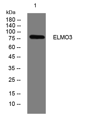
- Western blot analysis of lysates from CACO2 cells, primary antibody was diluted at 1:1000, 4°over night


