RAB3A rabbit pAb
- Catalog No.:YT7418
- Applications:WB;ELISA;IHC
- Reactivity:Human;Mouse;Rat
- Target:
- RAB3A
- Fields:
- >>Synaptic vesicle cycle;>>Insulin secretion
- Gene Name:
- RAB3A
- Protein Name:
- RAB3A
- Human Gene Id:
- 5864
- Human Swiss Prot No:
- P20336
- Mouse Gene Id:
- 19339
- Mouse Swiss Prot No:
- P63011
- Rat Gene Id:
- 25531
- Rat Swiss Prot No:
- P63012
- Immunogen:
- Synthesized peptide derived from human RAB3A AA range: 27-77
- Specificity:
- This antibody detects endogenous levels of RAB3A at Human/Mouse/Rat
- Formulation:
- Liquid in PBS containing 50% glycerol, 0.5% BSA and 0.02% sodium azide.
- Source:
- Polyclonal, Rabbit,IgG
- Dilution:
- WB 1:500-2000;IHC 1:50-300; ELISA 2000-20000
- Purification:
- The antibody was affinity-purified from rabbit antiserum by affinity-chromatography using epitope-specific immunogen.
- Concentration:
- 1 mg/ml
- Storage Stability:
- -15°C to -25°C/1 year(Do not lower than -25°C)
- Molecular Weight(Da):
- 24kD
- Function:
- function:Involved in exocytosis by regulating a late step in synaptic vesicle fusion. Could play a role in neurotransmitter release by regulating membrane flow in the nerve terminal.,similarity:Belongs to the small GTPase superfamily. Rab family.,subunit:Heterodimer with RIMS2. Part of a ternary complex involving PCLO and EPAC2. Interacts with RPH3A. Interacts with the exocyst complex through SEC15. Binds SYTL4, RIMS1 and RIMS2. Interacts with RAB3IP. Interacts with SGSM1 and SGSM3.,tissue specificity:Specifically expressed in brain.,
- Subcellular Location:
- Cytoplasm, cytosol . Lysosome . Cytoplasmic vesicle, secretory vesicle . Cell projection, axon . Cell membrane ; Lipid-anchor ; Cytoplasmic side . Cell junction, synapse, presynapse . Cell junction, synapse, postsynapse . Cycles between a vesicle-associated GTP-bound form and a cytosolic GDP-bound form. .
- Expression:
- Specifically expressed in brain.
- June 19-2018
- WESTERN IMMUNOBLOTTING PROTOCOL
- June 19-2018
- IMMUNOHISTOCHEMISTRY-PARAFFIN PROTOCOL
- June 19-2018
- IMMUNOFLUORESCENCE PROTOCOL
- September 08-2020
- FLOW-CYTOMEYRT-PROTOCOL
- May 20-2022
- Cell-Based ELISA│解您多样本WB检测之困扰
- July 13-2018
- CELL-BASED-ELISA-PROTOCOL-FOR-ACETYL-PROTEIN
- July 13-2018
- CELL-BASED-ELISA-PROTOCOL-FOR-PHOSPHO-PROTEIN
- July 13-2018
- Antibody-FAQs
- Products Images
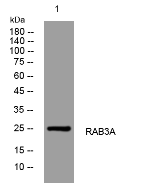
- Western blot analysis of lysates from HpeG2 cells, primary antibody was diluted at 1:1000, 4°over night
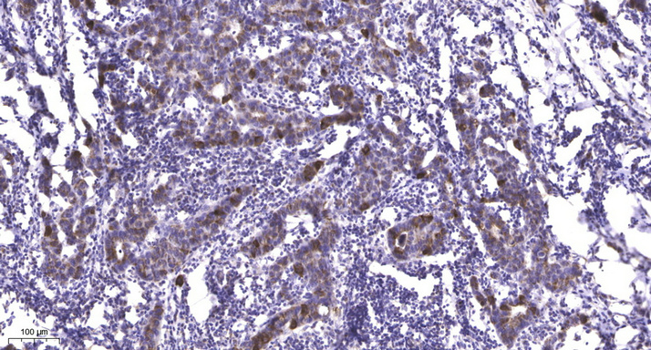
- Immunohistochemical analysis of paraffin-embedded human Breast cancer. 1, Antibody was diluted at 1:200(4° overnight). 2, Tris-EDTA,pH9.0 was used for antigen retrieval. 3,Secondary antibody was diluted at 1:200(room temperature, 45min).


