- Home
- About
- Promotions
-
Products
-
Elisa Kits
- |
-
Primary antibodies
- |
-
Secondary antibodies
- |
-
Proteins
- |
-
IHC reagents
- |
-
WB reagents
- PonceauS Staining Solution
- PBST Washing Buffer, 10X
- 1.5M Tris-HCl Buffer, pH8.8
- 1M Tris-HCl Buffer, pH6.8
- 10% SDS Solution
- Prestained Protein Marker
- TBST Washing Buffer, 10X
- SDS PAGE Loading Buffer, 5X
- Stripping Buffered Solution
- Tris Buffer, pH7.4, 10X
- Total Protein Extraction Kit
- Running Buffer, 10X
- Transfer Buffer, 10X
- 30% Acr-Bis(29:1) Solution
- Tris电泳液速溶颗粒
- PBS(1X, premixed powder)
- TBS(1X, premixed powder)
- 快速封闭液
- 转膜液速溶颗粒
- Chemical reagents
- News
- Distributor
- Resources
- Contact
- Home
- >
- Info
- >
- ARL6 rabbit pAb
- >
- Go Back
ARL6 rabbit pAb
- Catalog No.:YT7348
- Applications:WB
- Reactivity:Human;Mouse
- Immunogen:
- Synthesized peptide derived from human ARL6 AA range: 105-155
- Specificity:
- This antibody detects endogenous levels of ARL6 at Human/Mouse
- Formulation:
- Liquid in PBS containing 50% glycerol, 0.5% BSA and 0.02% sodium azide.
- Source:
- Polyclonal, Rabbit,IgG
- Purification:
- The antibody was affinity-purified from rabbit antiserum by affinity-chromatography using epitope-specific immunogen.
- Storage Stability:
- -15°C to -25°C/1 year(Do not lower than -25°C)
- Molecular Weight(Da):
- 20kD
- Background:
- The protein encoded by this gene belongs to the ARF-like (ADP ribosylation factor-like) sub-family of the ARF family of GTP-binding proteins which are involved in regulation of intracellular traffic. Mutations in this gene are associated with Bardet-Biedl syndrome (BBS). A vision-specific transcript, encoding long isoform BBS3L, has been described (PMID: 20333246). [provided by RefSeq, Apr 2016],
- Function:
- disease:Defects in ARL6 are a cause of Bardet-Biedl syndrome type 3 (BBS3) [MIM:209900]. Bardet-Biedl syndrome (BBS) is a genetically heterogeneous disorder characterized by usually severe pigmentary retinopathy, early onset obesity, polydactyly, hypogenitalism, renal malformation and mental retardation. Secondary features include diabetes mellitus, hypertension and congenital heart disease.,similarity:Belongs to the small GTPase superfamily. Arf family.,subunit:Interacts with SEC61B, ARL6IP1, ARL6IP2, ARL6IP3, ARL6IP4 ARL6IP5 and ARL6IP6.,
- Subcellular Location:
- Cell projection, cilium membrane; Peripheral membrane protein; Cytoplasmic side. Cytoplasm, cytoskeleton, cilium axoneme. Cytoplasm, cytoskeleton, cilium basal body. Appears in a pattern of punctae flanking the microtubule axoneme that likely correspond to small membrane-associated patches. Localizes to the so-called ciliary gate where vesicles carrying ciliary cargo fuse with the membrane.
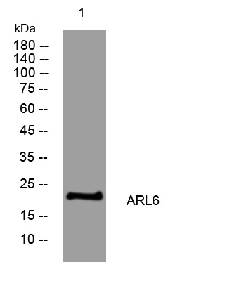
- Western blot analysis of lysates from MCF-7 cells, primary antibody was diluted at 1:1000, 4°over night


