- Home
- About
- Promotions
-
Products
-
Elisa Kits
- |
-
Primary antibodies
- |
-
Secondary antibodies
- |
-
Proteins
- |
-
IHC reagents
- |
-
WB reagents
- PonceauS Staining Solution
- PBST Washing Buffer, 10X
- 1.5M Tris-HCl Buffer, pH8.8
- 1M Tris-HCl Buffer, pH6.8
- 10% SDS Solution
- Prestained Protein Marker
- TBST Washing Buffer, 10X
- SDS PAGE Loading Buffer, 5X
- Stripping Buffered Solution
- Tris Buffer, pH7.4, 10X
- Total Protein Extraction Kit
- Running Buffer, 10X
- Transfer Buffer, 10X
- 30% Acr-Bis(29:1) Solution
- Tris电泳液速溶颗粒
- PBS(1X, premixed powder)
- TBS(1X, premixed powder)
- 快速封闭液
- 转膜液速溶颗粒
- Chemical reagents
- News
- Distributor
- Resources
- Contact
- Home
- >
- Info
- >
- TGM5 rabbit pAb
- >
- Go Back
TGM5 rabbit pAb
- Catalog No.:YT7219
- Applications:WB
- Reactivity:Human;Mouse
- Immunogen:
- Synthesized peptide derived from human TGM5 AA range: 448-498
- Specificity:
- This antibody detects endogenous levels of TGM5 at Human/Mouse
- Formulation:
- Liquid in PBS containing 50% glycerol, 0.5% BSA and 0.02% sodium azide.
- Source:
- Polyclonal, Rabbit,IgG
- Purification:
- The antibody was affinity-purified from rabbit antiserum by affinity-chromatography using epitope-specific immunogen.
- Storage Stability:
- -15°C to -25°C/1 year(Do not lower than -25°C)
- Molecular Weight(Da):
- 79kD
- Background:
- This gene encodes a member of the transglutaminase family. The encoded protein catalyzes formation of protein cross-links between glutamine and lysine residues, often resulting in stabilization of protein assemblies. This reaction is calcium dependent. Mutations in this gene have been associated with acral peeling skin syndrome. [provided by RefSeq, Oct 2009],
- Function:
- catalytic activity:Protein glutamine + alkylamine = protein N(5)-alkylglutamine + NH(3).,caution:The sequence shown here is derived from an Ensembl automatic analysis pipeline and should be considered as preliminary data.,cofactor:Binds 1 calcium ion per subunit.,disease:Defects in TGM5 are a cause of peeling skin syndrome acral type (APSS) [MIM:609796, 270300]. Peeling skin syndrome (PSS) is an autosomal recessive genodermatosis characterized by the continuous shedding of the outer layers of the epidermis from birth and throughout life. In some cases of PSS, skin peeling is accompanied by erythema, vesicular lesions, or, in rare cases, other ectodermal features, like fragile hair and nail abnormalities. Two main subtypes, noninflammatory type A and inflammatory type B, have been suggested. However, it is clear from the dermatology literature that there are additional subtypes. In some f
- Subcellular Location:
- Cytoplasm . Associated with intermediate filaments.
- Expression:
- Expressed in foreskin keratinocytes.
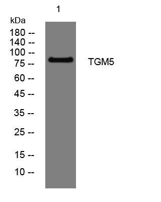
- Western blot analysis of lysates from U2OS cells, primary antibody was diluted at 1:1000, 4°over night


