- Home
- About
- Promotions
-
Products
-
Elisa Kits
- |
-
Primary antibodies
- |
-
Secondary antibodies
- |
-
Proteins
- |
-
IHC reagents
- |
-
WB reagents
- PonceauS Staining Solution
- PBST Washing Buffer, 10X
- 1.5M Tris-HCl Buffer, pH8.8
- 1M Tris-HCl Buffer, pH6.8
- 10% SDS Solution
- Prestained Protein Marker
- TBST Washing Buffer, 10X
- SDS PAGE Loading Buffer, 5X
- Stripping Buffered Solution
- Tris Buffer, pH7.4, 10X
- Total Protein Extraction Kit
- Running Buffer, 10X
- Transfer Buffer, 10X
- 30% Acr-Bis(29:1) Solution
- Tris电泳液速溶颗粒
- PBS(1X, premixed powder)
- TBS(1X, premixed powder)
- 快速封闭液
- 转膜液速溶颗粒
- Chemical reagents
- News
- Distributor
- Resources
- Contact
- Home
- >
- Info
- >
- F19A4 rabbit pAb
- >
- Go Back
F19A4 rabbit pAb
- Catalog No.:YT7208
- Applications:WB
- Reactivity:Human;Mouse
- Immunogen:
- Synthesized peptide derived from human F19A4 AA range: 40-90
- Specificity:
- This antibody detects endogenous levels of F19A4 at Human/Mouse
- Formulation:
- Liquid in PBS containing 50% glycerol, 0.5% BSA and 0.02% sodium azide.
- Source:
- Polyclonal, Rabbit,IgG
- Purification:
- The antibody was affinity-purified from rabbit antiserum by affinity-chromatography using epitope-specific immunogen.
- Storage Stability:
- -15°C to -25°C/1 year(Do not lower than -25°C)
- Molecular Weight(Da):
- 15kD
- Background:
- This gene is a member of the TAFA family which is composed of five highly homologous genes that encode small secreted proteins. These proteins contain conserved cysteine residues at fixed positions, and are distantly related to MIP-1alpha, a member of the CC-chemokine family. The TAFA proteins are predominantly expressed in specific regions of the brain, and are postulated to function as brain-specific chemokines or neurokines, that act as regulators of immune and nervous cells. Alternatively spliced transcript variants have been observed for this gene. [provided by RefSeq, Nov 2011],
- Function:
- caution:The sequence shown here is derived from an Ensembl automatic analysis pipeline and should be considered as preliminary data.,similarity:Belongs to the FAM19/TAFA family.,tissue specificity:Brain-specific.,
- Subcellular Location:
- Secreted .
- Expression:
- Expressed in brain (PubMed:15028294). Expressed in LPS-stimulated monocytes and macrophages, especially in polarized M1 (PubMed:25109685).
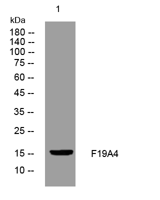
- Western blot analysis of lysates from CACO2 cells, primary antibody was diluted at 1:1000, 4°over night


