- Home
- About
- Promotions
-
Products
-
Elisa Kits
- |
-
Primary antibodies
- |
-
Secondary antibodies
- |
-
Proteins
- |
-
IHC reagents
- |
-
WB reagents
- PonceauS Staining Solution
- PBST Washing Buffer, 10X
- 1.5M Tris-HCl Buffer, pH8.8
- 1M Tris-HCl Buffer, pH6.8
- 10% SDS Solution
- Prestained Protein Marker
- TBST Washing Buffer, 10X
- SDS PAGE Loading Buffer, 5X
- Stripping Buffered Solution
- Tris Buffer, pH7.4, 10X
- Total Protein Extraction Kit
- Running Buffer, 10X
- Transfer Buffer, 10X
- 30% Acr-Bis(29:1) Solution
- Tris电泳液速溶颗粒
- PBS(1X, premixed powder)
- TBS(1X, premixed powder)
- 快速封闭液
- 转膜液速溶颗粒
- Chemical reagents
- News
- Distributor
- Resources
- Contact
- Home
- >
- Info
- >
- HERC3 rabbit pAb
- >
- Go Back
HERC3 rabbit pAb
- Catalog No.:YT7201
- Applications:WB
- Reactivity:Human
- Fields:
- >>Ubiquitin mediated proteolysis
- Gene Name:
- HERC3 KIAA0032
- Immunogen:
- Synthesized peptide derived from human HERC3 AA range: 985-1035
- Specificity:
- This antibody detects endogenous levels of HERC3 at Human
- Formulation:
- Liquid in PBS containing 50% glycerol, 0.5% BSA and 0.02% sodium azide.
- Source:
- Polyclonal, Rabbit,IgG
- Purification:
- The antibody was affinity-purified from rabbit antiserum by affinity-chromatography using epitope-specific immunogen.
- Storage Stability:
- -15°C to -25°C/1 year(Do not lower than -25°C)
- Molecular Weight(Da):
- 116kD
- Background:
- This gene encodes a member the HERC ubiquitin ligase family. The encoded protein is located in the cytosol and binds ubiquitin via a HECT domain. Mutations in this gene have been associated with colorectal and gastric carcinomas. Alternatively spliced transcript variants encoding multiple isoforms have been observed for this gene. [provided by RefSeq, Oct 2012],
- Function:
- function:E3 ubiquitin-protein ligase which accepts ubiquitin from an E2 ubiquitin-conjugating enzyme in the form of a thioester and then directly transfers the ubiquitin to targeted substrates.,pathway:Protein modification; protein ubiquitination.,PTM:Ubiquitinated; which promotes degradation by the proteasome.,similarity:Contains 1 HECT (E6AP-type E3 ubiquitin-protein ligase) domain.,similarity:Contains 7 RCC1 repeats.,subcellular location:Also found in vesicular-like structures.,
- Subcellular Location:
- Cytoplasm. Cytoplasmic vesicle. Also found in vesicular-like structures.
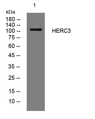
- Western blot analysis of lysates from 3T3 cells, primary antibody was diluted at 1:1000, 4°over night


