- Home
- About
- Promotions
-
Products
-
Elisa Kits
- |
-
Primary antibodies
- |
-
Secondary antibodies
- |
-
Proteins
- |
-
IHC reagents
- |
-
WB reagents
- PonceauS Staining Solution
- PBST Washing Buffer, 10X
- 1.5M Tris-HCl Buffer, pH8.8
- 1M Tris-HCl Buffer, pH6.8
- 10% SDS Solution
- Prestained Protein Marker
- TBST Washing Buffer, 10X
- SDS PAGE Loading Buffer, 5X
- Stripping Buffered Solution
- Tris Buffer, pH7.4, 10X
- Total Protein Extraction Kit
- Running Buffer, 10X
- Transfer Buffer, 10X
- 30% Acr-Bis(29:1) Solution
- Tris电泳液速溶颗粒
- PBS(1X, premixed powder)
- TBS(1X, premixed powder)
- 快速封闭液
- 转膜液速溶颗粒
- Chemical reagents
- News
- Distributor
- Resources
- Contact
- Home
- >
- Info
- >
- O10J4 rabbit pAb
- >
- Go Back
O10J4 rabbit pAb
- Catalog No.:YT6926
- Applications:WB
- Reactivity:Human
- Fields:
- >>Olfactory transduction
- Gene Name:
- OR10J4 OR10J4P
- Immunogen:
- Synthesized peptide derived from human O10J4 AA range: 102-152
- Specificity:
- This antibody detects endogenous levels of O10J4 at Human
- Formulation:
- Liquid in PBS containing 50% glycerol, 0.5% BSA and 0.02% sodium azide.
- Source:
- Polyclonal, Rabbit,IgG
- Purification:
- The antibody was affinity-purified from rabbit antiserum by affinity-chromatography using epitope-specific immunogen.
- Storage Stability:
- -15°C to -25°C/1 year(Do not lower than -25°C)
- Molecular Weight(Da):
- 34kD
- Background:
- Olfactory receptors interact with odorant molecules in the nose, to initiate a neuronal response that triggers the perception of a smell. The olfactory receptor proteins are members of a large family of G-protein-coupled receptors (GPCR) arising from single coding-exon genes. Olfactory receptors share a 7-transmembrane domain structure with many neurotransmitter and hormone receptors and are responsible for the recognition and G protein-mediated transduction of odorant signals. The olfactory receptor gene family is the largest in the genome. The nomenclature assigned to the olfactory receptor genes and proteins for this organism is independent of other organisms. [provided by RefSeq, Jul 2008],
- Function:
- function:Odorant receptor .,polymorphism:A single nucleotide deletion at position Ile-198 in the gene coding for this protein is reponsible for functional diversity thus producing a pseudogene.,similarity:Belongs to the G-protein coupled receptor 1 family.,
- Subcellular Location:
- Cell membrane; Multi-pass membrane protein.
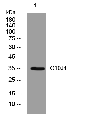
- Western blot analysis of lysates from MCF-7 cells, primary antibody was diluted at 1:1000, 4°over night


