- Home
- About
- Promotions
-
Products
-
Elisa Kits
- |
-
Primary antibodies
- |
-
Secondary antibodies
- |
-
Proteins
- |
-
IHC reagents
- |
-
WB reagents
- PonceauS Staining Solution
- PBST Washing Buffer, 10X
- 1.5M Tris-HCl Buffer, pH8.8
- 1M Tris-HCl Buffer, pH6.8
- 10% SDS Solution
- Prestained Protein Marker
- TBST Washing Buffer, 10X
- SDS PAGE Loading Buffer, 5X
- Stripping Buffered Solution
- Tris Buffer, pH7.4, 10X
- Total Protein Extraction Kit
- Running Buffer, 10X
- Transfer Buffer, 10X
- 30% Acr-Bis(29:1) Solution
- Tris电泳液速溶颗粒
- PBS(1X, premixed powder)
- TBS(1X, premixed powder)
- 快速封闭液
- 转膜液速溶颗粒
- Chemical reagents
- News
- Distributor
- Resources
- Contact
- Home
- >
- Info
- >
- CHP3 rabbit pAb
- >
- Go Back
CHP3 rabbit pAb
- Catalog No.:YT6823
- Applications:WB
- Reactivity:Human;Mouse
- Immunogen:
- Synthesized peptide derived from human CHP3 AA range: 20-70
- Specificity:
- This antibody detects endogenous levels of CHP3 at Human/Mouse
- Formulation:
- Liquid in PBS containing 50% glycerol, 0.5% BSA and 0.02% sodium azide.
- Source:
- Polyclonal, Rabbit,IgG
- Purification:
- The antibody was affinity-purified from rabbit antiserum by affinity-chromatography using epitope-specific immunogen.
- Storage Stability:
- -15°C to -25°C/1 year(Do not lower than -25°C)
- Molecular Weight(Da):
- 24kD
- Function:
- caution:It is uncertain whether Met-1 or Met-54 is the initiator.,cofactor:Binds calcium.,developmental stage:Strongly up-regulated in K562 cells treated by PMA to promote megakaryocytic differentiation, but not when treated by DMSO to promote granulocytic differentiation or by hemin to promote erythroid differentiation (at protein level).,function:Essential for the coupling of ERK cascade activation with the expression of ETS family genes in megakaryocytic differentiation.,similarity:Contains 1 EF-hand domain.,subunit:Interacts with Sodium/hydrogen exchanger 1 (SLC9A1).,tissue specificity:Abundantly expressed in heart.,
- Subcellular Location:
- Nucleus . Cytoplasm . Membrane ; Lipid-anchor . Cell membrane . Cell projection, lamellipodium . Cell projection, ruffle membrane . Colocalizes with SLC9A1 at the plasma membrane. .
- Expression:
- Expressed in mature megakaryocytes and polymorphonuclear granulocytes (at protein level). Abundantly expressed in heart. Also expressed at a lower level in adult testis and salivary gland, and in the placenta.
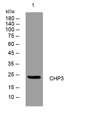
- Western blot analysis of lysates from HpeG2 cells, primary antibody was diluted at 1:1000, 4°over night


