PLCD3 rabbit pAb
- Catalog No.:YT6631
- Applications:WB;IHC
- Reactivity:Human;Mouse
- Target:
- PLCD3
- Fields:
- >>Inositol phosphate metabolism;>>Metabolic pathways;>>Calcium signaling pathway;>>Phosphatidylinositol signaling system;>>Thyroid hormone signaling pathway;>>AGE-RAGE signaling pathway in diabetic complications;>>Shigellosis
- Gene Name:
- PLCD3 KIAA1964
- Protein Name:
- PLCD3
- Human Gene Id:
- 113026
- Human Swiss Prot No:
- Q8N3E9
- Mouse Gene Id:
- 72469
- Mouse Swiss Prot No:
- Q8K2J0
- Immunogen:
- Synthesized peptide derived from human PLCD3 AA range: 501-551
- Specificity:
- This antibody detects endogenous levels of PLCD3 at Human/Mouse
- Formulation:
- Liquid in PBS containing 50% glycerol, 0.5% BSA and 0.02% sodium azide.
- Source:
- Polyclonal, Rabbit,IgG
- Dilution:
- WB 1:500-2000;IHC 1:50-300
- Purification:
- The antibody was affinity-purified from rabbit antiserum by affinity-chromatography using epitope-specific immunogen.
- Concentration:
- 1 mg/ml
- Storage Stability:
- -15°C to -25°C/1 year(Do not lower than -25°C)
- Molecular Weight(Da):
- 87kD
- Background:
- This gene encodes a member of the phospholipase C family, which catalyze the hydrolysis of phosphatidylinositol 4,5-bisphosphate to generate the second messengers diacylglycerol and inositol 1,4,5-trisphosphate (IP3). Diacylglycerol and IP3 mediate a variety of cellular responses to extracellular stimuli by inducing protein kinase C and increasing cytosolic Ca(2+) concentrations. This enzyme localizes to the plasma membrane and requires calcium for activation. Its activity is inhibited by spermine, sphingosine, and several phospholipids. [provided by RefSeq, Jul 2008],
- Function:
- catalytic activity:1-phosphatidyl-1D-myo-inositol 4,5-bisphosphate + H(2)O = 1D-myo-inositol 1,4,5-trisphosphate + diacylglycerol.,cofactor:Binds 3 calcium ions per subunit. Two of the calcium ions are bound to the C2 domain.,domain:The C2 domain is a Ca(2+)-dependent membrane-targeting module.,domain:The PH domain mediates interaction with the surface membrane by binding to PIP2.,enzyme regulation:Strongly activated by phosphatidic acid. Inhibited by phosphatidylethanolamine (PtdEtn), phosphatidylcholine (PtdCho), sphingomyelin and phosphatidylserine (PtdSer).,function:Hydrolyzes the phosphatidylinositol 4,5-bisphosphate (PIP2) to generate 2 second messenger molecules diacylglycerol (DAG) and inositol 1,4,5-trisphosphate (IP3). DAG mediates the activation of protein kinase C (PKC), while IP3 releases Ca(2+) from intracellular stores. Essential for trophoblast and placental development.
- Subcellular Location:
- Membrane; Peripheral membrane protein. Cytoplasm. Cleavage furrow . Localizes at the cleavage furrow during cytokinesis. .
- Expression:
- Present in corneal epithelial cells (at protein level).
- June 19-2018
- WESTERN IMMUNOBLOTTING PROTOCOL
- June 19-2018
- IMMUNOHISTOCHEMISTRY-PARAFFIN PROTOCOL
- June 19-2018
- IMMUNOFLUORESCENCE PROTOCOL
- September 08-2020
- FLOW-CYTOMEYRT-PROTOCOL
- May 20-2022
- Cell-Based ELISA│解您多样本WB检测之困扰
- July 13-2018
- CELL-BASED-ELISA-PROTOCOL-FOR-ACETYL-PROTEIN
- July 13-2018
- CELL-BASED-ELISA-PROTOCOL-FOR-PHOSPHO-PROTEIN
- July 13-2018
- Antibody-FAQs
- Products Images
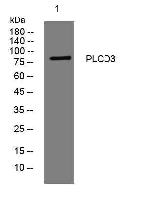
- Western blot analysis of lysates from MCF-7 cells, primary antibody was diluted at 1:1000, 4°over night
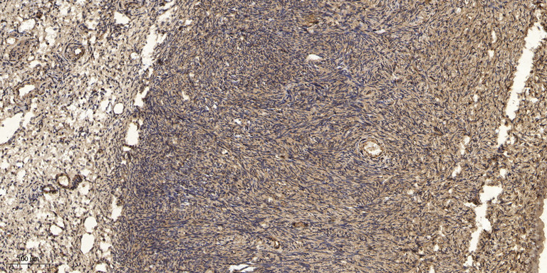
- Immunohistochemical analysis of paraffin-embedded human oophoroma. 1, Antibody was diluted at 1:200(4° overnight). 2, Tris-EDTA,pH9.0 was used for antigen retrieval. 3,Secondary antibody was diluted at 1:200(room temperature, 45min).


