FCGRN rabbit pAb
- Catalog No.:YT6440
- Applications:WB
- Reactivity:Human;Mouse;Rat
- Target:
- FCGRN
- Gene Name:
- FCGRT FCRN
- Protein Name:
- FCGRN
- Human Gene Id:
- 2217
- Human Swiss Prot No:
- P55899
- Mouse Gene Id:
- 14132
- Mouse Swiss Prot No:
- Q61559
- Rat Gene Id:
- 29558
- Rat Swiss Prot No:
- P13599
- Immunogen:
- Synthesized peptide derived from human FCGRN AA range: 146-196
- Specificity:
- This antibody detects endogenous levels of FCGRN at Human/Mouse/Rat
- Formulation:
- Liquid in PBS containing 50% glycerol, 0.5% BSA and 0.02% sodium azide.
- Source:
- Polyclonal, Rabbit,IgG
- Dilution:
- WB 1:500-2000
- Purification:
- The antibody was affinity-purified from rabbit antiserum by affinity-chromatography using epitope-specific immunogen.
- Concentration:
- 1 mg/ml
- Storage Stability:
- -15°C to -25°C/1 year(Do not lower than -25°C)
- Molecular Weight(Da):
- 40kD
- Background:
- This gene encodes a receptor that binds the Fc region of monomeric immunoglobulin G. The encoded protein transfers immunoglobulin G antibodies from mother to fetus across the placenta. This protein also binds immunoglobulin G to protect the antibody from degradation. Alternative splicing results in multiple transcript variants. [provided by RefSeq, Apr 2009],
- Function:
- function:Binds to the Fc region of monomeric immunoglobulins gamma. Mediates the uptake of IgG from milk. Possible role in transfer of immunoglobulin G from mother to fetus.,similarity:Belongs to the immunoglobulin superfamily.,subunit:FcRn complex consist of two subunits: p51, and p14 which is equivalent to beta-2-microglobulin. It forms an MHC class I-like heterodimer.,
- Subcellular Location:
- Cell membrane ; Single-pass type I membrane protein . Endosome membrane .
- Expression:
- Expressed in full-term placenta, heart, lung, liver, muscle, kidney, pancreas, and both fetal and adult small intestine.
- June 19-2018
- WESTERN IMMUNOBLOTTING PROTOCOL
- June 19-2018
- IMMUNOHISTOCHEMISTRY-PARAFFIN PROTOCOL
- June 19-2018
- IMMUNOFLUORESCENCE PROTOCOL
- September 08-2020
- FLOW-CYTOMEYRT-PROTOCOL
- May 20-2022
- Cell-Based ELISA│解您多样本WB检测之困扰
- July 13-2018
- CELL-BASED-ELISA-PROTOCOL-FOR-ACETYL-PROTEIN
- July 13-2018
- CELL-BASED-ELISA-PROTOCOL-FOR-PHOSPHO-PROTEIN
- July 13-2018
- Antibody-FAQs
- Products Images
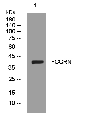
- Western blot analysis of lysates from Hela cells, primary antibody was diluted at 1:1000, 4°over night


