DEGS2 rabbit pAb
- Catalog No.:YT6346
- Applications:WB
- Reactivity:Human;Mouse;Rat
- Target:
- DEGS2
- Fields:
- >>Sphingolipid metabolism;>>Metabolic pathways;>>Sphingolipid signaling pathway
- Gene Name:
- DEGS2 C14orf66
- Protein Name:
- DEGS2
- Human Gene Id:
- 123099
- Human Swiss Prot No:
- Q6QHC5
- Mouse Gene Id:
- 70059
- Mouse Swiss Prot No:
- Q8R2F2
- Rat Gene Id:
- 314438
- Rat Swiss Prot No:
- Q564G3
- Immunogen:
- Synthesized peptide derived from human DEGS2 AA range: 92-142
- Specificity:
- This antibody detects endogenous levels of DEGS2 at Human/Mouse/Rat
- Formulation:
- Liquid in PBS containing 50% glycerol, 0.5% BSA and 0.02% sodium azide.
- Source:
- Polyclonal, Rabbit,IgG
- Dilution:
- WB 1:500-2000
- Purification:
- The antibody was affinity-purified from rabbit antiserum by affinity-chromatography using epitope-specific immunogen.
- Concentration:
- 1 mg/ml
- Storage Stability:
- -15°C to -25°C/1 year(Do not lower than -25°C)
- Molecular Weight(Da):
- 36kD
- Background:
- This gene encodes a bifunctional enzyme that is involved in the biosynthesis of phytosphingolipids in human skin and in other phytosphingolipid-containing tissues. This enzyme can act as a sphingolipid delta(4)-desaturase, and also as a sphingolipid C4-hydroxylase. [provided by RefSeq, Oct 2008],
- Function:
- function:Bifunctional enzyme which acts as both a sphingolipid delta(4)-desaturase and a sphingolipid C4-hydroxylase.,induction:Up-regulated during keratinocyte differentiation. Not expressed at day 0 or day 3 after differentiation, detected on day 6 and increases by day 9.,pathway:Membrane lipid metabolism; sphingolipid biosynthesis.,similarity:Belongs to the fatty acid desaturase family. DEGS subfamily.,tissue specificity:Highly expressed in skin, intestine and kidney.,
- Subcellular Location:
- Endoplasmic reticulum membrane ; Multi-pass membrane protein .
- Expression:
- Highly expressed in skin, intestine and kidney.
- June 19-2018
- WESTERN IMMUNOBLOTTING PROTOCOL
- June 19-2018
- IMMUNOHISTOCHEMISTRY-PARAFFIN PROTOCOL
- June 19-2018
- IMMUNOFLUORESCENCE PROTOCOL
- September 08-2020
- FLOW-CYTOMEYRT-PROTOCOL
- May 20-2022
- Cell-Based ELISA│解您多样本WB检测之困扰
- July 13-2018
- CELL-BASED-ELISA-PROTOCOL-FOR-ACETYL-PROTEIN
- July 13-2018
- CELL-BASED-ELISA-PROTOCOL-FOR-PHOSPHO-PROTEIN
- July 13-2018
- Antibody-FAQs
- Products Images
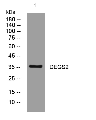
- Western blot analysis of lysates from MCF-7 cells, primary antibody was diluted at 1:1000, 4°over night


