Caspase-8 Polyclonal Antibody
- Catalog No.:YT6191
- Applications:IHC;IF;WB
- Reactivity:Human;Mouse;Rat;Pig;Chicken
- Target:
- Caspase-8
- Fields:
- >>Platinum drug resistance;>>p53 signaling pathway;>>Apoptosis;>>Apoptosis - multiple species;>>Necroptosis;>>Toll-like receptor signaling pathway;>>NOD-like receptor signaling pathway;>>RIG-I-like receptor signaling pathway;>>C-type lectin receptor signaling pathway;>>IL-17 signaling pathway;>>TNF signaling pathway;>>Non-alcoholic fatty liver disease;>>Alcoholic liver disease;>>Alzheimer disease;>>Huntington disease;>>Pathways of neurodegeneration - multiple diseases;>>Pathogenic Escherichia coli infection;>>Salmonella infection;>>Legionellosis;>>Chagas disease;>>Toxoplasmosis;>>Tuberculosis;>>Hepatitis C;>>Hepatitis B;>>Measles;>>Human cytomegalovirus infection;>>Influenza A;>>Human papillomavirus infection;>>Kaposi sarcoma-associated herpesvirus infection;>>Herpes simplex virus 1 infection;>>Epstein-Barr virus infection;>>Human immunodeficiency virus 1 infection;>>Pathways in cancer;>>Viral carcinogenesis;>>Viral myocarditis;>>Lipid and atherosclerosis
- Gene Name:
- CASP8 MCH5
- Protein Name:
- Caspase8
- Human Gene Id:
- 841
- Human Swiss Prot No:
- Q14790
- Immunogen:
- Synthesized peptide derived from human Caspase-8
- Specificity:
- This antibody detects endogenous levels of human Caspase-8
- Formulation:
- Liquid in PBS containing 50% glycerol, 0.5% BSA and 0.02% sodium azide.
- Source:
- Polyclonal, Rabbit,IgG
- Dilution:
- IHC 1:50-200, WB 1:500-2000. IF 1:50-200
- Purification:
- The antibody was affinity-purified from rabbit antiserum by affinity-chromatography using epitope-specific immunogen.
- Concentration:
- 1 mg/ml
- Storage Stability:
- -15°C to -25°C/1 year(Do not lower than -25°C)
- Other Name:
- Caspase-8 (CASP-8;EC 3.4.22.61;Apoptotic cysteine protease;Apoptotic protease Mch-5;CAP4;FADD-homologous ICE/ced-3-like protease;FADD-like ICE;FLICE;ICE-like apoptotic protease 5;MORT1-associated ced-3 homolog;MACH) [Cleaved into: Caspase-8 subunit p18;Caspase-8 subunit p10]
- Observed Band(KD):
- 55kD
- Background:
- This gene encodes a member of the cysteine-aspartic acid protease (caspase) family. Sequential activation of caspases plays a central role in the execution-phase of cell apoptosis. Caspases exist as inactive proenzymes composed of a prodomain, a large protease subunit, and a small protease subunit. Activation of caspases requires proteolytic processing at conserved internal aspartic residues to generate a heterodimeric enzyme consisting of the large and small subunits. This protein is involved in the programmed cell death induced by Fas and various apoptotic stimuli. The N-terminal FADD-like death effector domain of this protein suggests that it may interact with Fas-interacting protein FADD. This protein was detected in the insoluble fraction of the affected brain region from Huntington disease patients but not in those from normal controls, which implicated the role in neurodegenerative diseases. Many alt
- Function:
- catalytic activity:Strict requirement for Asp at position P1 and has a preferred cleavage sequence of (Leu/Asp/Val)-Glu-Thr-Asp-|-(Gly/Ser/Ala).,disease:Defects in CASP8 are the cause of caspase-8 deficiency (CASP8D) [MIM:607271]. CASP8D is a disorder resembling autoimmune lymphoproliferative syndrome (ALPS). It is characterized by lymphadenopathy, splenomegaly, and defective CD95-induced apoptosis of peripheral blood lymphocytes (PBLs). It leads to defects in activation of T-lymphocytes, B-lymphocytes, and natural killer cells leading to immunodeficiency characterized by recurrent sinopulmonary and herpes simplex virus infections and poor responses to immunization.,domain:Isoform 9 contains a N-terminal extension that is required for interaction with the BCAP31 complex.,function:Most upstream protease of the activation cascade of caspases responsible for the TNFRSF6/FAS mediated and TNF
- Subcellular Location:
- Cytoplasm . Nucleus .
- Expression:
- Isoform 1, isoform 5 and isoform 7 are expressed in a wide variety of tissues. Highest expression in peripheral blood leukocytes, spleen, thymus and liver. Barely detectable in brain, testis and skeletal muscle.
Effect of Trimethyltin chloride on proliferation and cell cycle of intestinal porcine epithelial cells. COMPARATIVE BIOCHEMISTRY AND PHYSIOLOGY C-TOXICOLOGY & PHARMACOLOGY Comp Biochem Phys C. 2021 Nov;249:109131 WB Pig Intestinal porcine epithelial (IPEC-J2) cell
Ganoderic Acids Prevent Renal Ischemia Reperfusion Injury by Inhibiting Inflammation and Apoptosis. INTERNATIONAL JOURNAL OF MOLECULAR SCIENCES Int J Mol Sci. 2021 Jan;22(19):10229 WB Rat Renal tissues NRK-52E cell
The role of Se content in improving anti-tumor activities and its potential mechanism for selenized Artemisia sphaerocephala polysaccharides. Food & Function Food Funct. 2021 Mar;12(5):2058-2074 WB Human HepG2 cell
SPAG6 silencing inhibits the growth of the malignant myeloid cell lines SKM-1 and K562 via activating p53 and caspase activation-dependent apoptosis. INTERNATIONAL JOURNAL OF ONCOLOGY 2015 Feb 01 WB Human 1:500 SKM-1 cell, K562 cell
Heme oxygenase-1 (HO-1) protects human lens epithelial cells (SRA01/04) against hydrogen peroxide (H2O2)-induced oxidative stress and apoptosis. EXPERIMENTAL EYE RESEARCH 2016 Mar 16 WB Human 1:1000 LEC cell
Anti-apoptotic and apoptotic pathway analysis of arsenic trioxide‑induced apoptosis in human gastric cancer SGC-7901 cells. ONCOLOGY REPORTS 2014 Jun 20 WB Human 1:800 SGC-7901 cell
miR-2954 Inhibits PI3K Signaling and Induces Autophagy and Apoptosis in Myocardium Selenium Deficiency. CELLULAR PHYSIOLOGY AND BIOCHEMISTRY 2018 Nov 21 WB Chicken 1:500 cardiomyocytes
Overexpression of miR‐200b inhibits the cell proliferation and promotes apoptosis of human hypertrophic scar fibroblasts in vitro. JOURNAL OF DERMATOLOGY WB Human hHSFs cell
The preparation of a cold-water soluble polysaccharide from Grifola frondosa and its inhibitory effects on MKN-45 cells. GLYCOCONJUGATE JOURNAL 2020 Jun 15 WB Human MKN-45 cell
Fucoidan inhibits proliferation of the SKM-1 acute myeloid leukaemia cell line via the activation of apoptotic pathways and production of reactive oxygen species. Molecular Medicine Reports Mol Med Rep. 2015 Nov;12(5):6649-6655 WB Human SKM-1 cell
A novel monoclonal antibody associated with glucoside kills gastric adenocarcinoma AGS cells based on glycosylation target JOURNAL OF CELLULAR AND MOLECULAR MEDICINE Heng Xu WB Human
The outer membrane protein Tp92 of Treponema pallidum delays human neutrophil apoptosis via the ERK, PI3K/Akt, and NF-κB pathways. MOLECULAR MICROBIOLOGY Yimou Wu WB Human human polymorphonuclear neutrophils (hPMNs)
- June 19-2018
- WESTERN IMMUNOBLOTTING PROTOCOL
- June 19-2018
- IMMUNOHISTOCHEMISTRY-PARAFFIN PROTOCOL
- June 19-2018
- IMMUNOFLUORESCENCE PROTOCOL
- September 08-2020
- FLOW-CYTOMEYRT-PROTOCOL
- May 20-2022
- Cell-Based ELISA│解您多样本WB检测之困扰
- July 13-2018
- CELL-BASED-ELISA-PROTOCOL-FOR-ACETYL-PROTEIN
- July 13-2018
- CELL-BASED-ELISA-PROTOCOL-FOR-PHOSPHO-PROTEIN
- July 13-2018
- Antibody-FAQs
- Products Images

- 57 Yang B, Wang L, Luo X, et al. SPAG6 silencing inhibits the growth of the malignant myeloid cell lines SKM-1 and K562 via activating p53 and caspase activation-dependent apoptosis[J]. International journal of oncology, 2015, 46(2): 649-656.
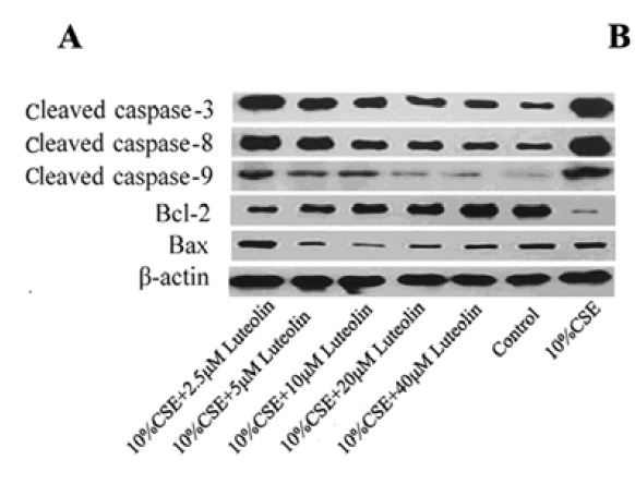
- 536 Tan X, Jin P, Feng L, et al. Protective effect of luteolin on cigarette smoke extract‑induced cellular toxicity and apoptosis in normal human bronchial epithelial cells via the Nrf2 pathway[J]. Oncology reports, 2014, 31(4): 1855-1862.

- 801 Li P, He Q Y, Luo C Q. Overexpression of miR‐200b inhibits the cell proliferation and promotes apoptosis of human hypertrophic scar fibroblasts in vitro[J]. The Journal of dermatology, 2014, 41(10): 903-911.

- 860 Yu Y, Yang Y, Wang J. Anti-apoptotic and apoptotic pathway analysis of arsenic trioxide‑induced apoptosis in human gastric cancer SGC-7901 cells[J]. Oncology reports, 2014, 32(3): 973-978.
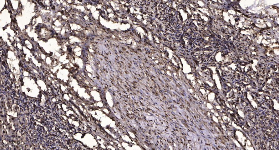
- Immunohistochemical analysis of paraffin-embedded human liver cancer. 1, Antibody was diluted at 1:200(4° overnight). 2, Tris-EDTA,pH9.0 was used for antigen retrieval. 3,Secondary antibody was diluted at 1:200(room temperature, 45min).


