COL8A2 Polyclonal Antibody
- Catalog No.:YT6088
- Applications:WB;ELISA
- Reactivity:Human;Mouse;Rat
- Target:
- COL8A2
- Fields:
- >>Protein digestion and absorption
- Gene Name:
- COL8A2
- Protein Name:
- COL8A2
- Human Gene Id:
- 1296
- Human Swiss Prot No:
- P25067
- Mouse Gene Id:
- 329941
- Mouse Swiss Prot No:
- P25318
- Immunogen:
- Synthesized peptide derived from human COL8A2. at AA range: 611-660
- Specificity:
- COL8A2 Polyclonal Antibody detects endogenous levels of COL8A2
- Formulation:
- Liquid in PBS containing 50% glycerol, 0.5% BSA and 0.02% sodium azide.
- Source:
- Polyclonal, Rabbit,IgG
- Dilution:
- WB 1:500-2000, ELISA 1:10000-20000
- Purification:
- The antibody was affinity-purified from rabbit antiserum by affinity-chromatography using epitope-specific immunogen.
- Concentration:
- 1 mg/ml
- Storage Stability:
- -15°C to -25°C/1 year(Do not lower than -25°C)
- Other Name:
- Collagen alpha-2(VIII) chain (Endothelial collagen)
- Observed Band(KD):
- 80kD
- Background:
- This gene encodes the alpha 2 chain of type VIII collagen. This protein is a major component of the basement membrane of the corneal endothelium and forms homo- or heterotrimers with alpha 1 (VIII) type collagens. Defects in this gene are associated with Fuchs endothelial corneal dystrophy and posterior polymorphous corneal dystrophy type 2. Alternative splicing results in multiple transcript variants. [provided by RefSeq, Jun 2014],
- Function:
- disease:Defects in COL8A2 are a cause of Fuchs endothelial corneal dystrophy (FECD) [MIM:136800]. FECD is the commonest primary disorder of the corneal endothelium in developed countries. Symptoms of painful visual loss result from corneal decompensation. Signs may be present from the fourth decade of life onwards. Tipically, focal wart-like guttata arising from Descemet membrane develops in the central cornea; Descemet membrane is thickened by abnormal collagenous deposition. FECD is usually sporadic but familial highly penetrant forms showing autosomal dominant inheritance are also recognized.,disease:Defects in COL8A2 are a cause of posterior polymorphous corneal dystrophy (PPCD) [MIM:122000]. PPCD is a slowly progressive hereditary disorder of the corneal endothelium that leads to a variable degree of visual impairment usually in adulthood. PPCD is usually inherited as an autosomal d
- Subcellular Location:
- Secreted, extracellular space, extracellular matrix, basement membrane.
- Expression:
- Expressed primarily in the subendothelium of large blood vessels. Also expressed in arterioles and venules in muscle, heart, kidney, spleen, umbilical cord, liver and lung and is also found in connective tissue layers around hair follicles, around nerve bundles in muscle, in the dura of the optic nerve, in cornea and sclera, and in the perichondrium of cartilaginous tissues. In the kidney, expressed in mesangial cells, glomerular endothelial cells, and tubular epithelial cells. Also expressed in mast cells, and in astrocytes during the repair process. Expressed in Descemet's membrane.
- June 19-2018
- WESTERN IMMUNOBLOTTING PROTOCOL
- June 19-2018
- IMMUNOHISTOCHEMISTRY-PARAFFIN PROTOCOL
- June 19-2018
- IMMUNOFLUORESCENCE PROTOCOL
- September 08-2020
- FLOW-CYTOMEYRT-PROTOCOL
- May 20-2022
- Cell-Based ELISA│解您多样本WB检测之困扰
- July 13-2018
- CELL-BASED-ELISA-PROTOCOL-FOR-ACETYL-PROTEIN
- July 13-2018
- CELL-BASED-ELISA-PROTOCOL-FOR-PHOSPHO-PROTEIN
- July 13-2018
- Antibody-FAQs
- Products Images
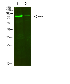
- Western Blot analysis of 1,hela 2,3T3 cells using primary antibody diluted at 1:500(4°C overnight). Secondary antibody:Goat Anti-rabbit IgG IRDye 800( diluted at 1:5000, 25°C, 1 hour)


