Caspase-1 Polyclonal Antibody
- Catalog No.:YT5743
- Applications:IF;WB;IHC;ELISA
- Reactivity:Human;Mouse;Rat
- Target:
- Caspase-1
- Fields:
- >>Necroptosis;>>Neutrophil extracellular trap formation;>>NOD-like receptor signaling pathway;>>Cytosolic DNA-sensing pathway;>>C-type lectin receptor signaling pathway;>>Amyotrophic lateral sclerosis;>>Pathogenic Escherichia coli infection;>>Shigellosis;>>Salmonella infection;>>Pertussis;>>Legionellosis;>>Yersinia infection;>>Influenza A;>>Coronavirus disease - COVID-19;>>Lipid and atherosclerosis
- Gene Name:
- CASP1 IL1BC IL1BCE
- Protein Name:
- Caspase1
- Human Gene Id:
- 834
- Human Swiss Prot No:
- P29466
- Mouse Gene Id:
- 12362
- Mouse Swiss Prot No:
- P29452
- Rat Swiss Prot No:
- P43527
- Immunogen:
- The antiserum was produced against synthesized peptide derived from the C-terminal region of human CASP1. AA range:350-400
- Specificity:
- Caspase-1 Polyclonal Antibody detects endogenous levels of Caspase-1
- Formulation:
- Liquid in PBS containing 50% glycerol, 0.5% BSA and 0.02% sodium azide.
- Source:
- Polyclonal, Rabbit,IgG
- Dilution:
- IF 1:50-200 WB 1:500-2000, IHC 1:50-300, ELISA 1:10000-20000
- Purification:
- The antibody was affinity-purified from rabbit antiserum by affinity-chromatography using epitope-specific immunogen.
- Concentration:
- 1 mg/ml
- Storage Stability:
- -15°C to -25°C/1 year(Do not lower than -25°C)
- Other Name:
- caspase 1, apoptosis-related cysteine peptidase (interleukin 1, beta, convertase)
- Molecular Weight(Da):
- 45kD
- Observed Band(KD):
- 45kD, 35kD, cleaced isform p10 :10kD
- Background:
- This gene encodes a protein which is a member of the cysteine-aspartic acid protease (caspase) family. Sequential activation of caspases plays a central role in the execution-phase of cell apoptosis. Caspases exist as inactive proenzymes which undergo proteolytic processing at conserved aspartic residues to produce 2 subunits, large and small, that dimerize to form the active enzyme. This gene was identified by its ability to proteolytically cleave and activate the inactive precursor of interleukin-1, a cytokine involved in the processes such as inflammation, septic shock, and wound healing. This gene has been shown to induce cell apoptosis and may function in various developmental stages. Studies of a similar gene in mouse suggest a role in the pathogenesis of Huntington disease. Alternative splicing results in transcript variants encoding distinct isoforms. [provided by RefSeq, Mar 2012],
- Function:
- alternative products:Additional isoforms seem to exist,catalytic activity:Strict requirement for an Asp residue at position P1 and has a preferred cleavage sequence of Tyr-Val-Ala-Asp-|-.,enzyme regulation:Specifically inhibited by the cowpox virus Crma protein.,function:Thiol protease that cleaves IL-1 beta between an Asp and an Ala, releasing the mature cytokine which is involved in a variety of inflammatory processes. Important for defense against pathogens. Cleaves and activates sterol regulatory element binding proteins (SREBPs). Can also promote apoptosis.,PTM:The two subunits are derived from the precursor sequence by an autocatalytic mechanism.,similarity:Belongs to the peptidase C14A family.,similarity:Contains 1 CARD domain.,subunit:Heterotetramer that consists of two anti-parallel arranged heterodimers, each one formed by a 20 kDa (p20) and a 10 kDa (p10) subunit. The p20 subu
- Subcellular Location:
- Cytoplasm . Cell membrane .
- Expression:
- Expressed in larger amounts in spleen and lung. Detected in liver, heart, small intestine, colon, thymus, prostate, skeletal muscle, peripheral blood leukocytes, kidney and testis. No expression in the brain.
Platelet-rich plasma-derived exosomes attenuate intervertebral disc degeneration by promoting NLRP3 autophagic degradation in macrophages WB Rat 1:1000 /BMDMs
Ras guanine nucleotide-releasing protein-4 promotes renal inflammatory injury in type 2 diabetes mellitus Metabolism. 2022 Feb;:155177. WB Mouse Renal
Crocin alleviates cognitive impairment associated with atherosclerosis via improving neuroinflammation in LDLR−/− mice fed a high-fat/cholesterol diet 2022 Jan 27. WB Mouse 1:1000 hippocampus,parietal cortex
Effects of Hypoxic Environment on Periodontal Tissue through the ROS/TXNIP/NLRP3 Inflammasome Pathway Biomed Res Int. 2022;2022:7690960. WB,IF,IHC Rat ,Human 1 : 1000,1 : 250,1 : 250 Periodontal
Dl-3-n-butylphthalide inhibits neuroinflammation by stimulating foxp3 and Ki-67 in an ischemic stroke model.. Aging-US Aging-Us. 2021 Feb 15; 13(3): 3763–3778 IHC Rat 1:50 Brain
Folic Acid Alleviates High Glucose and Fat-Induced Pyroptosis via Inhibition of the Hippo Signal Pathway on H9C2 Cells. Frontiers in Molecular Biosciences Front Mol Biosci. 2021; 8: 698698 WB Mouse 1:1000 Heart H9C2 cell
Neuroprotective Effects of Safflower Flavonoid Extract in 6-Hydroxydopamine-Induced Model of Parkinson’s Disease May Be Related to its Anti-Inflammatory Action. MOLECULES Molecules. 2020 Jan;25(21):5206 WB Mouse 1:1000 Brain(substantia nigra)
Cereal Fiber Ameliorates High-Fat/Cholesterol-Diet-Induced Atherosclerosis by Modulating the NLRP3 Inflammasome Pathway in ApoE–/– Mice. JOURNAL OF AGRICULTURAL AND FOOD CHEMISTRY J Agr Food Chem. 2018;66(19):4827–4834 WB Mouse 1:1000 aortas
Oat fiber supplementation alleviates intestinal inflammation and ameliorates intestinal mucosal barrier via acting on gut microbiota-derived metabolites in LDLR–/– mice. NUTRITION Nutrition. 2022 Mar;95:111558 WB Mouse Aortic tissue,intestinal tissue
Antitumor Effect of Fluoxetine on Chronic Stress-Promoted Lung Cancer Growth via Suppressing Kynurenine Pathway and Enhancing Cellular Immunity. Frontiers in Pharmacology Front Pharmacol. 2021 Aug;0:1609 WB Mouse 1:2000 A549 cell-xenograft
Apple Polyphenols Extract (APE) Alleviated Dextran Sulfate Sodium Induced Acute Ulcerative Colitis and Accompanying Neuroinflammation via Inhibition of Apoptosis and Pyroptosis. Foods Foods. 2021 Nov;10(11):2711 WB Mouse Colon
MCC950 Inhibits NLRP3 Inflammasome and Alleviates Axonal Injures in Early Stages of Diffuse Axonal Injury in Rats. NEUROCHEMICAL RESEARCH Neurochem Res. 2020 Sep;45(9):2020-2031 WB Rat 1:1000 Cortex
Early changes of NLRP3 inflammasome activation after hypoxic-ischemic brain injury in neonatal rats. International Journal of Clinical and Experimental Pathology Int J Clin Exp Patho. 2021; 14(2): 209–220 IHC Rat 1 : 500 Brain
Liu, Xi, et al. "Antineuroinflammatory effects of dl-3-n-butylphthalide conferred by stimulation of Foxp3 and Ki-67 in an ischemic stroke model." (2020).
Gynostemma pentaphyllum polysaccharides ameliorate non-alcoholic steatohepatitis in mice associated with gut microbiota and the TLR2/NLRP3 pathway Frontiers in Endocrinology Rui-Rui Wang WB Mouse
Puerarin protects against sepsis-associated encephalopathy by inhibiting NLRP3/Caspase-1/GSDMD pyroptosis pathway and reducing blood-brain barrier damage EUROPEAN JOURNAL OF PHARMACOLOGY Min Zhao IHC Mouse,Rat brain tissues
VX-765 ameliorates CKD VSMC calcification by regulating STAT3 activation EUROPEAN JOURNAL OF PHARMACOLOGY Zhangxiu He IHC,IF Rat abdominal aorta Primary rat VSMCs (rVSMCs)
Hyperhomocysteinemia activates NLRP3 inflammasome to cause hepatic steatosis and insulin resistance via MDM2-mediated ubiquitination of HSF1 INTERNATIONAL IMMUNOPHARMACOLOGY Cheng Zeng IF,WB Mouse,Human liver tissue HepG2 cell
Glucose-driven transformable complex eliminates biofilm and alleviates inflamm-aging for diabetic periodontitis therapy. Bin Liu WB Mouse 1:1500 RAW264.7 cell
Induced Neural Stem Cells Suppressed Neuroinflammation by Inhibiting the Microglial Pyroptotic Pathway in Intracerebral Hemorrhage Rats. Xueyun Liang IF Rat,Human 1:300 brain tissue induced neural stem cells (iNSCs)
Phloretin ameliorates heart function after myocardial infarction via NLRP3/Caspase-1/IL-1β signaling. Xuejun Jiang WB Rat 1:1000 cardiac tissue H9c2 Cell
Phloretin ameliorates heart function after myocardial infarction via NLRP3/Caspase-1/IL-1β signaling. Xuejun Jiang WB Rat 1:1000 cardiac tissue H9c2 Cell
Targeting the mechanism of IRF3 in sepsis-associated acute kidney injury via the Hippo pathway. Xingui Dai WB Human HK-2 cell
A novel drug combination of Tofacitinib and Iguratimod alleviates rheumatoid arthritis and secondary osteoporosis. INTERNATIONAL IMMUNOPHARMACOLOGY Qiang Shu IHC Rat 1:100 Synovial tissue,bone tissue
ERRα promotes glycolytic metabolism and targets the NLRP3/caspase-1/GSDMD pathway to regulate pyroptosis in endometrial cancer. JOURNAL OF EXPERIMENTAL & CLINICAL CANCER RESEARCH Pengming Sun WB Human KLE cell,HEC-1A cell
Piceatannol alleviates liver ischaemia/reperfusion injury by inhibiting TLR4/NF-κB/NLRP3 in hepatic macrophages. EUROPEAN JOURNAL OF PHARMACOLOGY Jian Du IHC,IF,WB Mouse 1:50,1:50,1:1000 liver tissue macrophages
Huangqi Baihe Granules alleviate hypobaric hypoxia-induced acute lung injury in rats by suppressing oxidative stress and the TLR4/NF-κB/NLRP3 inflammatory pathway JOURNAL OF ETHNOPHARMACOLOGY Yuanding Zeng WB Rat 1:1000 lung tissue
Blocking CIRP protects against acute pancreatitis by improving mitochondrial function and suppressing pyroptosis in acinar cells Cell Death Discovery Liu Wuming WB Mouse,Rat 1:1000 pancreatic tissue AR42J cell
Dihydroartemisinin-driven TOM70 inhibition leads to mitochondrial destabilization to induce pyroptosis against lung cancer PHYTOTHERAPY RESEARCH Liu-Gen Li WB,IHC Mouse,Human Lewis cell-Xenograft Lewis cell,A549 cell
Targeting TANK-binding kinase 1 attenuates painful diabetic neuropathy via inhibiting microglia pyroptosis Cell Communication and Signaling Liao Qinming WB Mouse spinal cord tissue
Icariin suppresses nephrotic syndrome by inhibiting pyroptosis and epithelial-to-mesenchymal transition PLoS One Shuwen Duan WB Rat renal tissue
- June 19-2018
- WESTERN IMMUNOBLOTTING PROTOCOL
- June 19-2018
- IMMUNOHISTOCHEMISTRY-PARAFFIN PROTOCOL
- June 19-2018
- IMMUNOFLUORESCENCE PROTOCOL
- September 08-2020
- FLOW-CYTOMEYRT-PROTOCOL
- May 20-2022
- Cell-Based ELISA│解您多样本WB检测之困扰
- July 13-2018
- CELL-BASED-ELISA-PROTOCOL-FOR-ACETYL-PROTEIN
- July 13-2018
- CELL-BASED-ELISA-PROTOCOL-FOR-PHOSPHO-PROTEIN
- July 13-2018
- Antibody-FAQs
- Products Images
- Immunofluorescence analysis of A549. 1,primary Antibody(red) was diluted at 1:200(4°C overnight). 2, Goat Anti Rabbit IgG (H&L) - Alexa Fluor 594 Secondary antibody was diluted at 1:1000(room temperature, 50min).3, Picture B: DAPI(blue) 10min.
.jpg)
- Liu, Xi et al. “Dl-3-n-butylphthalide inhibits neuroinflammation by stimulating foxp3 and Ki-67 in an ischemic stroke model.” Agingvol. 13,3 (2021): 3763-3778. doi:10.18632/aging.202338

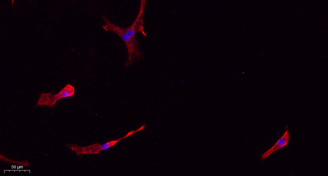
- Immunofluorescence analysis of rat-lung tissue. 1,Caspase-1 Polyclonal Antibody(red) was diluted at 1:200(4°C,overnight). 2, Cy3 labled Secondary antibody was diluted at 1:300(room temperature, 50min).3, Picture B: DAPI(blue) 10min. Picture A:Target. Picture B: DAPI. Picture C: merge of A+B

- Immunofluorescence analysis of rat-lung tissue. 1,Caspase-1 Polyclonal Antibody(red) was diluted at 1:200(4°C,overnight). 2, Cy3 labled Secondary antibody was diluted at 1:300(room temperature, 50min).3, Picture B: DAPI(blue) 10min. Picture A:Target. Picture B: DAPI. Picture C: merge of A+B
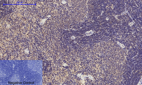
- Immunohistochemical analysis of paraffin-embedded Rat-spleen tissue. 1,Caspase-1 Polyclonal Antibody was diluted at 1:200(4°C,overnight). 2, Sodium citrate pH 6.0 was used for antibody retrieval(>98°C,20min). 3,Secondary antibody was diluted at 1:200(room tempeRature, 30min). Negative control was used by secondary antibody only.
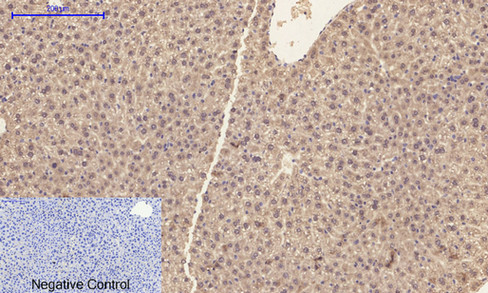
- Immunohistochemical analysis of paraffin-embedded Mouse-liver tissue. 1,Caspase-1 Polyclonal Antibody was diluted at 1:200(4°C,overnight). 2, Sodium citrate pH 6.0 was used for antibody retrieval(>98°C,20min). 3,Secondary antibody was diluted at 1:200(room tempeRature, 30min). Negative control was used by secondary antibody only.
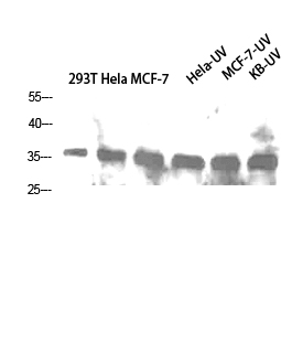
- Western Blot analysis of 293T Hela MCF-7 Hela-UV MCF-7-UV KB-UV cells using Caspase-1 Polyclonal Antibody diluted at 1:1000. Secondary antibody(catalog#:RS0002) was diluted at 1:20000
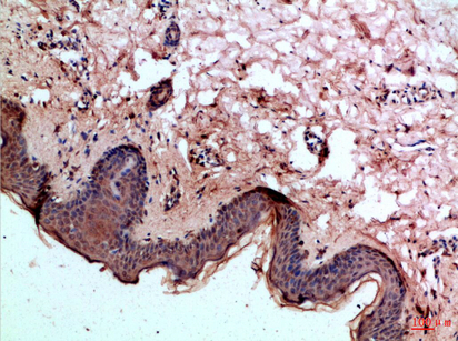
- Immunohistochemical analysis of paraffin-embedded Human-skin, antibody was diluted at 1:100
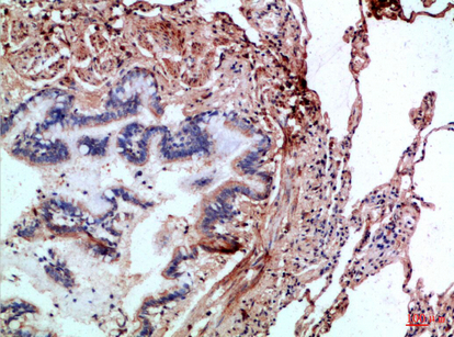
- Immunohistochemical analysis of paraffin-embedded Human-lung, antibody was diluted at 1:100
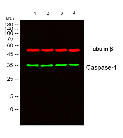
- Western blot analysis of lysates from 1) 293T , 2) Hela ,3) MCF-7, 4) Hela-UV cells, (Green) primary antibody was diluted at 1:1000, 4°over night, secondary antibody(cat:RS23920)was diluted at 1:10000, 37° 1hour. (Red) Tubulin β Monoclonal Antibody(5G3) (cat:YM3030) antibody was diluted at 1:5000 as loading control, 4° over night,secondary antibody(cat:RS23710)was diluted at 1:10000, 37° 1hour.


