CD158e Polyclonal Antibody
- Catalog No.:YT5716
- Applications:WB;IHC;IF;ELISA
- Reactivity:Human;Rat;Mouse;
- Target:
- CD158e
- Fields:
- >>Antigen processing and presentation;>>Natural killer cell mediated cytotoxicity;>>Graft-versus-host disease
- Gene Name:
- KIR3DL1
- Protein Name:
- Killer cell immunoglobulin-like receptor 3DL1
- Human Gene Id:
- 3811
- Human Swiss Prot No:
- P43629
- Mouse Swiss Prot No:
- P83555
- Rat Gene Id:
- 353253
- Rat Swiss Prot No:
- P83556
- Immunogen:
- Synthesized peptide derived from Killer cell immunoglobulin-like receptor 3DL1 at AA range: 21-70
- Specificity:
- CD158e Polyclonal Antibody detects endogenous levels of CD158e protein.
- Formulation:
- Liquid in PBS containing 50% glycerol, 0.5% BSA and 0.02% sodium azide.
- Source:
- Polyclonal, Rabbit,IgG
- Dilution:
- WB 1:500 - 1:2000. IHC: 1:100-1:300. ELISA: 1:10000.. IF 1:50-200
- Purification:
- The antibody was affinity-purified from rabbit antiserum by affinity-chromatography using epitope-specific immunogen.
- Concentration:
- 1 mg/ml
- Storage Stability:
- -15°C to -25°C/1 year(Do not lower than -25°C)
- Other Name:
- KIR3DL1;CD158E;NKAT3;NKB1;Killer cell immunoglobulin-like receptor 3DL1;CD158 antigen-like family member E;HLA-BW4-specific inhibitory NK cell receptor;MHC class I NK cell receptor;Natural killer-associated transcript 3;NKAT-3;p70 natural killer cell receptor clones CL-2/CL-11;p70 NK receptor CL-2/CL-11;CD158e
- Observed Band(KD):
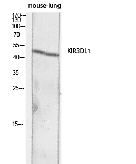
- 50kD
- Background:
- killer cell immunoglobulin like receptor, three Ig domains and long cytoplasmic tail 1(KIR3DL1) Homo sapiens Killer cell immunoglobulin-like receptors (KIRs) are transmembrane glycoproteins expressed by natural killer cells and subsets of T cells. The KIR genes are polymorphic and highly homologous and they are found in a cluster on chromosome 19q13.4 within the 1 Mb leukocyte receptor complex (LRC). The gene content of the KIR gene cluster varies among haplotypes, although several "framework" genes are found in all haplotypes (KIR3DL3, KIR3DP1, KIR3DL4, KIR3DL2). The KIR proteins are classified by the number of extracellular immunoglobulin domains (2D or 3D) and by whether they have a long (L) or short (S) cytoplasmic domain. KIR proteins with the long cytoplasmic domain transduce inhibitory signals upon ligand binding via an immune tyrosine-based inhibitory motif (ITIM), while KIR proteins with the short cytoplasmic domain lack the
- Function:
- function:Receptor on natural killer (NK) cells for HLA Bw4 allele. Inhibits the activity of NK cells thus preventing cell lysis.,function:Receptor on natural killer (NK) cells for HLA-C alleles. Does not inhibit the activity of NK cells.,polymorphism:The KIR genes are located in a segment of DNA on 19q13.4 in the leukocyte receptor complex that has undergone expansion and contraction over time, probably through unequal crossing-over. Thus, KIR haplotypes vary in the number and types of genes, although a few framework loci, such as the gene KIR3DL1, are present on all or nearly all haplotypes. KIR3DL1 and KIR3DS1 segregate as alleles of the locus KIR3DL1/3DS1.,similarity:Belongs to the immunoglobulin superfamily.,similarity:Contains 3 Ig-like C2-type (immunoglobulin-like) domains.,tissue specificity:Expressed in NK and T-cell lines but not in B-lymphoblastoid cell lines or in a colon carc
- Subcellular Location:
- Cell membrane; Single-pass type I membrane protein.
- Expression:
- Blood,Brain,Lymphoi
- June 19-2018
- WESTERN IMMUNOBLOTTING PROTOCOL
- June 19-2018
- IMMUNOHISTOCHEMISTRY-PARAFFIN PROTOCOL
- June 19-2018
- IMMUNOFLUORESCENCE PROTOCOL
- September 08-2020
- FLOW-CYTOMEYRT-PROTOCOL
- May 20-2022
- Cell-Based ELISA│解您多样本WB检测之困扰
- July 13-2018
- CELL-BASED-ELISA-PROTOCOL-FOR-ACETYL-PROTEIN
- July 13-2018
- CELL-BASED-ELISA-PROTOCOL-FOR-PHOSPHO-PROTEIN
- July 13-2018
- Antibody-FAQs
- Products Images

- Western blot analysis of mouse-lung lysis using KIR3DL1 antibody. Antibody was diluted at 1:1000. Secondary antibody(catalog#:RS0002) was diluted at 1:20000

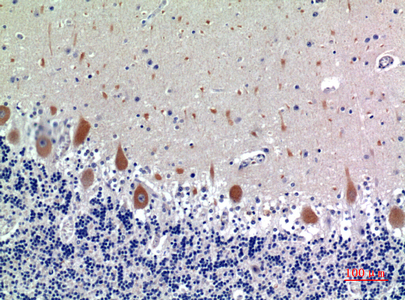
- Immunohistochemical analysis of paraffin-embedded human-brain, antibody was diluted at 1:100

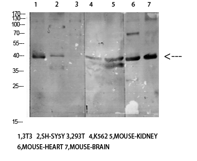
- Western Blot analysis of various cells using Antibody diluted at 1:1000. Secondary antibody(catalog#:RS0002) was diluted at 1:20000


