BST-2 Polyclonal Antibody
- Catalog No.:YT5612
- Applications:WB;IHC;IF;ELISA
- Reactivity:Human;Rat;Mouse;
- Target:
- BST-2
- Fields:
- >>Viral life cycle - HIV-1;>>Herpes simplex virus 1 infection;>>Human immunodeficiency virus 1 infection
- Gene Name:
- BST2
- Protein Name:
- Bone marrow stromal antigen 2
- Human Gene Id:
- 684
- Human Swiss Prot No:
- Q10589
- Mouse Swiss Prot No:
- Q8R2Q8
- Immunogen:
- The antiserum was produced against synthesized peptide derived from the Internal region of human BST2. AA range:101-150
- Specificity:
- BST-2 Polyclonal Antibody detects endogenous levels of BST-2 protein.
- Formulation:
- Liquid in PBS containing 50% glycerol, 0.5% BSA and 0.02% sodium azide.
- Source:
- Polyclonal, Rabbit,IgG
- Dilution:
- WB 1:500 - 1:2000. IHC: 1:100-1:300. ELISA: 1:10000.. IF 1:50-200
- Purification:
- The antibody was affinity-purified from rabbit antiserum by affinity-chromatography using epitope-specific immunogen.
- Concentration:
- 1 mg/ml
- Storage Stability:
- -15°C to -25°C/1 year(Do not lower than -25°C)
- Other Name:
- BST2;Bone marrow stromal antigen 2;BST-2;HM1.24 antigen;Tetherin;CD317
- Observed Band(KD):
- 20kD
- Background:
- Bone marrow stromal cells are involved in the growth and development of B-cells. The specific function of the protein encoded by the bone marrow stromal cell antigen 2 is undetermined; however, this protein may play a role in pre-B-cell growth and in rheumatoid arthritis. [provided by RefSeq, Jul 2008],
- Function:
- disease:May play a role in B-cell activation in rheumatoid arthritis (RA).,function:May be involved in the sorting of secreted proteins (By similarity). May be involved in pre-B-cell growth. Antiretroviral defense protein, that blocks release of retrovirus from the cell surface. Depleted unpon HIV-1 infection by viral VPU protein through 20S proteasome degradation.,induction:During B-cell activation (at protein level), or by interferon alpha as part of entiviral state cellular program.,subunit:Homodimer.,tissue specificity:Predominantly expressed in liver, lung, heart and placenta. Lower levels in pancreas, kidney, skeletal muscle and brain. Overexpressed in multiple myeloma cells. Highly expressed during B-cell development, from pro-B precursors to plasma cells. Highy expressed on T-cells, monocytes, NK cells and dendritic cells (at protein level).,
- Subcellular Location:
- Golgi apparatus, trans-Golgi network. Cell membrane ; Single-pass type II membrane protein. Cell membrane ; Lipid-anchor, GPI-anchor . Membrane raft. Cytoplasm. Apical cell membrane . Shuttles between the cell membrane, where it is present predominantly in membrane/lipid rafts, and the trans-Golgi network. Forms a complex with MMP14 and localizes to the cytoplasm.; Golgi apparatus, trans-Golgi network . Late endosome . (Microbial infection) HIV-1 VPU and HIV-2 ENV can target it to the trans-Golgi network thus sequestering it away from virus assembly sites on the cell membrane. Targeted to late endosomes upon KSHV infection and subsequent ubiquitination. .
- Expression:
- Predominantly expressed in liver, lung, heart and placenta. Lower levels in pancreas, kidney, skeletal muscle and brain. Overexpressed in multiple myeloma cells. Highly expressed during B-cell development, from pro-B precursors to plasma cells. Highly expressed on T-cells, monocytes, NK cells and dendritic cells (at protein level).
- June 19-2018
- WESTERN IMMUNOBLOTTING PROTOCOL
- June 19-2018
- IMMUNOHISTOCHEMISTRY-PARAFFIN PROTOCOL
- June 19-2018
- IMMUNOFLUORESCENCE PROTOCOL
- September 08-2020
- FLOW-CYTOMEYRT-PROTOCOL
- May 20-2022
- Cell-Based ELISA│解您多样本WB检测之困扰
- July 13-2018
- CELL-BASED-ELISA-PROTOCOL-FOR-ACETYL-PROTEIN
- July 13-2018
- CELL-BASED-ELISA-PROTOCOL-FOR-PHOSPHO-PROTEIN
- July 13-2018
- Antibody-FAQs
- Products Images
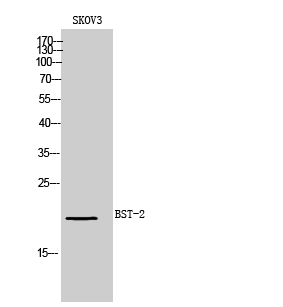
- Western Blot analysis of SKOV3 cells using BST-2 Polyclonal Antibody. Secondary antibody(catalog#:RS0002) was diluted at 1:20000
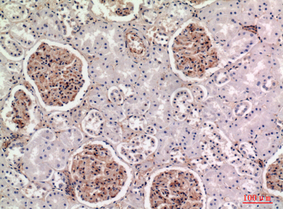
- Immunohistochemical analysis of paraffin-embedded human-kidney, antibody was diluted at 1:100


