Jagged1 Polyclonal Antibody
- Catalog No.:YT5401
- Applications:IF;WB;IHC;ELISA
- Reactivity:Human;Mouse;Rat
- Target:
- Jagged1
- Fields:
- >>Endocrine resistance;>>Notch signaling pathway;>>Apelin signaling pathway;>>Th1 and Th2 cell differentiation;>>TNF signaling pathway;>>Human papillomavirus infection;>>Pathways in cancer;>>Chemical carcinogenesis - receptor activation;>>Breast cancer
- Gene Name:
- JAG1
- Protein Name:
- Protein jagged-1
- Human Gene Id:
- 182
- Human Swiss Prot No:
- P78504
- Mouse Gene Id:
- 16449
- Mouse Swiss Prot No:
- Q9QXX0
- Rat Gene Id:
- 29146
- Rat Swiss Prot No:
- Q63722
- Immunogen:
- The antiserum was produced against synthesized peptide derived from the Internal region of human JAG1. AA range:981-1030
- Specificity:
- Jagged1 Polyclonal Antibody detects endogenous levels of Jagged1 protein.
- Formulation:
- Liquid in PBS containing 50% glycerol, 0.5% BSA and 0.02% sodium azide.
- Source:
- Polyclonal, Rabbit,IgG
- Dilution:
- IF 1:50-200 WB 1:500 - 1:2000. IHC: 1:100-1:300. ELISA: 1:20000. Not yet tested in other applications.
- Purification:
- The antibody was affinity-purified from rabbit antiserum by affinity-chromatography using epitope-specific immunogen.
- Concentration:
- 1 mg/ml
- Storage Stability:
- -15°C to -25°C/1 year(Do not lower than -25°C)
- Other Name:
- JAG1;JAGL1;Protein jagged-1;Jagged1;hJ1;CD339
- Observed Band(KD):
- 140kD
- Background:
- The jagged 1 protein encoded by JAG1 is the human homolog of the Drosophilia jagged protein. Human jagged 1 is the ligand for the receptor notch 1, the latter a human homolog of the Drosophilia jagged receptor notch. Mutations that alter the jagged 1 protein cause Alagille syndrome. Jagged 1 signalling through notch 1 has also been shown to play a role in hematopoiesis. [provided by RefSeq, Jul 2008],
- Function:
- developmental stage:Expressed in 32-52 days embryos in the distal cardiac outflow tract and pulmonary artery, major arteries, portal vein, optic vesicle, otocyst, branchial arches, metanephros, pancreas, mesocardium, around the major bronchial branches, and in the neural tube.,disease:Defects in JAG1 are a cause of tetralogy of Fallot (TOF) [MIM:187500]. TOF is a congenital heart anomaly which consists of pulmonary stenosis, ventricular septal defect, dextroposition of the aorta (aorta is on the right side instead of the left) and hypertrophy of the right ventricle. This condition results in a blue baby at birth due to inadequate oxygenation. Surgical correction is emergent.,disease:Defects in JAG1 are the cause of Alagille syndrome type 1 (ALGS1) [MIM:118450]. Alagille syndrome is an autosomal dominant multisystem disorder defined clinically by hepatic bile duct paucity and cholestasis
- Subcellular Location:
- Membrane; Single-pass type I membrane protein.
- Expression:
- Widely expressed in adult and fetal tissues. In cervix epithelium expressed in undifferentiated subcolumnar reserve cells and squamous metaplasia. Expression is up-regulated in cervical squamous cell carcinoma. Expressed in bone marrow cell line HS-27a which supports the long-term maintenance of immature progenitor cells.
Endosulfan triggers epithelial-mesenchymal transition via PTP4A3-mediated TGF-β signaling pathway in prostate cancer cells. SCIENCE OF THE TOTAL ENVIRONMENT Sci Total Environ. 2020 Aug;731:139234 WB Human PC3 cell, DU145 cells
Downregulation of miR-19a exhibits inhibitory effects on metastatic renal cell carcinoma by targeting PIK3CA and inactivating Notch signaling in vitro. ONCOLOGY REPORTS Oncol Rep. 2015 Aug;34(2):739-746 WB Human 1:100 786-O cell, Caki-1 cell
Endothelin-1 Activates the Notch Signaling Pathway and Promotes Tumorigenesis in Giant Cell Tumor of the Spine. SPINE Spine. 2019 Sep;44(17):E1000-E1009 WB Human 1:500 giant cell tumor (GCT) tissue
Role of the Notch ligands Jagged1 and Delta4 in Th17/Treg immune imbalance in a mouse model of chronic asthma. EXPERIMENTAL LUNG RESEARCH Exp Lung Res. 2021;47(6):289-299 WB Mouse Lung
- June 19-2018
- WESTERN IMMUNOBLOTTING PROTOCOL
- June 19-2018
- IMMUNOHISTOCHEMISTRY-PARAFFIN PROTOCOL
- June 19-2018
- IMMUNOFLUORESCENCE PROTOCOL
- September 08-2020
- FLOW-CYTOMEYRT-PROTOCOL
- May 20-2022
- Cell-Based ELISA│解您多样本WB检测之困扰
- July 13-2018
- CELL-BASED-ELISA-PROTOCOL-FOR-ACETYL-PROTEIN
- July 13-2018
- CELL-BASED-ELISA-PROTOCOL-FOR-PHOSPHO-PROTEIN
- July 13-2018
- Antibody-FAQs
- Products Images

- Immunofluorescence analysis of A549. 1,primary Antibody(red) was diluted at 1:200(4°C overnight). 2, Goat Anti Rabbit IgG (H&L) - Alexa Fluor 594 Secondary antibody was diluted at 1:1000(room temperature, 50min).3, Picture B: DAPI(blue) 10min.
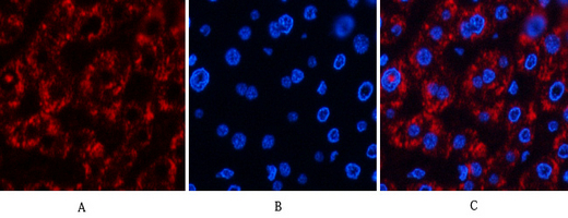
- Immunofluorescence analysis of human-liver tissue. 1,Jagged1 Polyclonal Antibody(red) was diluted at 1:200(4°C,overnight). 2, Cy3 labled Secondary antibody was diluted at 1:300(room temperature, 50min).3, Picture B: DAPI(blue) 10min. Picture A:Target. Picture B: DAPI. Picture C: merge of A+B
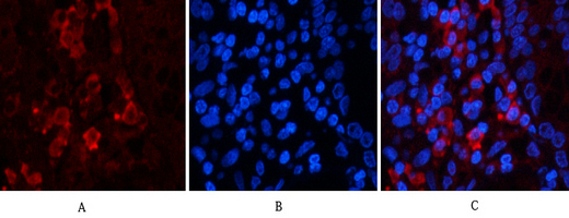
- Immunofluorescence analysis of human-stomach tissue. 1,Jagged1 Polyclonal Antibody(red) was diluted at 1:200(4°C,overnight). 2, Cy3 labled Secondary antibody was diluted at 1:300(room temperature, 50min).3, Picture B: DAPI(blue) 10min. Picture A:Target. Picture B: DAPI. Picture C: merge of A+B
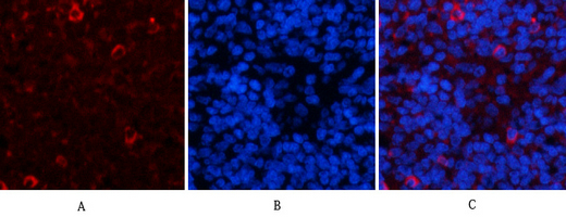
- Immunofluorescence analysis of rat-spleen tissue. 1,Jagged1 Polyclonal Antibody(red) was diluted at 1:200(4°C,overnight). 2, Cy3 labled Secondary antibody was diluted at 1:300(room temperature, 50min).3, Picture B: DAPI(blue) 10min. Picture A:Target. Picture B: DAPI. Picture C: merge of A+B
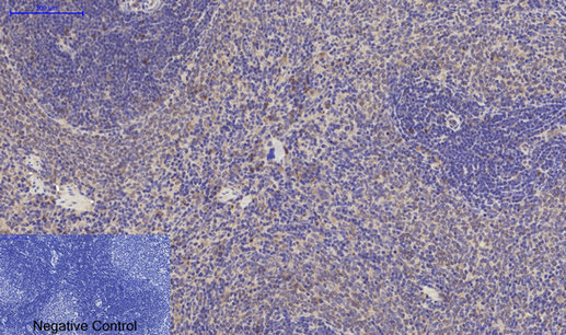
- Immunohistochemical analysis of paraffin-embedded Rat-spleen tissue. 1,Jagged1 Polyclonal Antibody was diluted at 1:200(4°C,overnight). 2, Sodium citrate pH 6.0 was used for antibody retrieval(>98°C,20min). 3,Secondary antibody was diluted at 1:200(room tempeRature, 30min). Negative control was used by secondary antibody only.
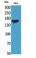
- Western Blot analysis of HeLa cells using Jagged1 Polyclonal Antibody. Secondary antibody(catalog#:RS0002) was diluted at 1:20000
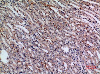
- Immunohistochemical analysis of paraffin-embedded mouse-kidney, antibody was diluted at 1:100
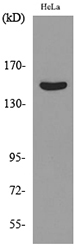
- Western blot analysis of lysate from HeLa cells, using JAG1 Antibody.


