Laminin γ-2 Polyclonal Antibody
- Catalog No.:YT5379
- Applications:WB;IHC;IF;ELISA
- Reactivity:Human;Rat
- Target:
- Laminin γ-2
- Fields:
- >>PI3K-Akt signaling pathway;>>Focal adhesion;>>ECM-receptor interaction;>>Toxoplasmosis;>>Amoebiasis;>>Human papillomavirus infection;>>Pathways in cancer;>>Small cell lung cancer
- Gene Name:
- LAMC2
- Protein Name:
- Laminin subunit gamma-2
- Human Gene Id:
- 3918
- Human Swiss Prot No:
- Q13753
- Mouse Swiss Prot No:
- Q61092
- Immunogen:
- The antiserum was produced against synthesized peptide derived from the C-terminal region of human LAMC2. AA range:1021-1070
- Specificity:
- Laminin γ-2 Polyclonal Antibody detects endogenous levels of Laminin γ-2 protein.
- Formulation:
- Liquid in PBS containing 50% glycerol, 0.5% BSA and 0.02% sodium azide.
- Source:
- Polyclonal, Rabbit,IgG
- Dilution:
- WB 1:500 - 1:2000. IHC: 1:100-1:300. ELISA: 1:20000.. IF 1:50-200
- Purification:
- The antibody was affinity-purified from rabbit antiserum by affinity-chromatography using epitope-specific immunogen.
- Concentration:
- 1 mg/ml
- Storage Stability:
- -15°C to -25°C/1 year(Do not lower than -25°C)
- Other Name:
- LAMC2;LAMB2T;LAMNB2;Laminin subunit gamma-2;Cell-scattering factor 140 kDa subunit;CSF 140 kDa subunit;Epiligrin subunit gamma;Kalinin subunit gamma;Kalinin/nicein/epiligrin 100 kDa subunitLadsin 140 kDa subunit;Laminin B2t chain;Laminin-5 subunit gamma;Large adhesive scatter factor 140 kDa subunit;Nicein subunit gamma
- Observed Band(KD):
- 135kD
- Background:
- Laminins, a family of extracellular matrix glycoproteins, are the major noncollagenous constituent of basement membranes. They have been implicated in a wide variety of biological processes including cell adhesion, differentiation, migration, signaling, neurite outgrowth and metastasis. Laminins, composed of 3 non identical chains: laminin alpha, beta and gamma (formerly A, B1, and B2, respectively), have a cruciform structure consisting of 3 short arms, each formed by a different chain, and a long arm composed of all 3 chains. Each laminin chain is a multidomain protein encoded by a distinct gene. Several isoforms of each chain have been described. Different alpha, beta and gamma chain isomers combine to give rise to different heterotrimeric laminin isoforms which are designated by Arabic numerals in the order of their discovery, i.e. alpha1beta1gamma1 heterotrimer is laminin 1. The biological func
- Function:
- disease:Defects in LAMC2 are a cause of epidermolysis bullosa junctional Herlitz type (H-JEB) [MIM:226700]; also known as junctional epidermolysis bullosa Herlitz-Pearson type. JEB defines a group of blistering skin diseases characterized by tissue separation which occurs within the dermo-epidermal basement membrane. H-JEB is a severe, infantile and lethal form. Death occurs usually within the first six months of life. Occasionally, children survive to teens. H-JEB is marked by bullous lesions at birth and extensive denudation of skin and mucous membranes that may be hemorrhagic.,domain:Domain IV is globular.,domain:The alpha-helical domains I and II are thought to interact with other laminin chains to form a coiled coil structure.,function:Binding to cells via a high affinity receptor, laminin is thought to mediate the attachment, migration and organization of cells into tissues during
- Subcellular Location:
- Secreted, extracellular space, extracellular matrix, basement membrane. Major component.
- Expression:
- The large variant is expressed only in specific epithelial cells of embryonic and neonatal tissues. In 17-week old embryo the small variant is found in cerebral cortex, lung, and distal tubes of kidney, but not in epithelia except for distal tubuli.
Capsaicin and Cold exposure promote EMT-mediated premetastatic niche formation to facilitate colorectal cancer metastasis Journal of Cancer Feifei Nong IHC,WB Rat 1:50,1:1000 colon tissue
- June 19-2018
- WESTERN IMMUNOBLOTTING PROTOCOL
- June 19-2018
- IMMUNOHISTOCHEMISTRY-PARAFFIN PROTOCOL
- June 19-2018
- IMMUNOFLUORESCENCE PROTOCOL
- September 08-2020
- FLOW-CYTOMEYRT-PROTOCOL
- May 20-2022
- Cell-Based ELISA│解您多样本WB检测之困扰
- July 13-2018
- CELL-BASED-ELISA-PROTOCOL-FOR-ACETYL-PROTEIN
- July 13-2018
- CELL-BASED-ELISA-PROTOCOL-FOR-PHOSPHO-PROTEIN
- July 13-2018
- Antibody-FAQs
- Products Images
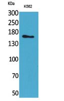
- Western Blot analysis of K562 cells using Laminin γ-2 Polyclonal Antibody. Secondary antibody(catalog#:RS0002) was diluted at 1:20000
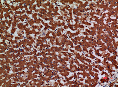
- Immunohistochemical analysis of paraffin-embedded human-liver, antibody was diluted at 1:100
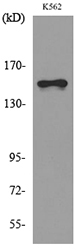
- Western blot analysis of lysate from K562 cells, using LAMC2 Antibody.


