CDCP1 Polyclonal Antibody
- Catalog No.:YT5291
- Applications:WB;IHC;IF;ELISA
- Reactivity:Human;Mouse;Rat
- Target:
- CDCP1
- Gene Name:
- CDCP1
- Protein Name:
- CUB domain-containing protein 1
- Human Gene Id:
- 64866
- Human Swiss Prot No:
- Q9H5V8
- Mouse Gene Id:
- 109332
- Mouse Swiss Prot No:
- Q5U462
- Immunogen:
- The antiserum was produced against synthesized peptide derived from the Internal region of human CDCP1. AA range:241-290
- Specificity:
- CDCP1 Polyclonal Antibody detects endogenous levels of CDCP1 protein.
- Formulation:
- Liquid in PBS containing 50% glycerol, 0.5% BSA and 0.02% sodium azide.
- Source:
- Polyclonal, Rabbit,IgG
- Dilution:
- WB 1:500 - 1:2000. IHC: 1:100-300 ELISA: 1:20000.. IF 1:50-200
- Purification:
- The antibody was affinity-purified from rabbit antiserum by affinity-chromatography using epitope-specific immunogen.
- Concentration:
- 1 mg/ml
- Storage Stability:
- -15°C to -25°C/1 year(Do not lower than -25°C)
- Other Name:
- CDCP1;TRASK;CUB domain-containing protein 1;Membrane glycoprotein gp140;Subtractive immunization M plus HEp3-associated 135 kDa protein;SIMA135;Transmembrane and associated with src kinases;CD318
- Observed Band(KD):
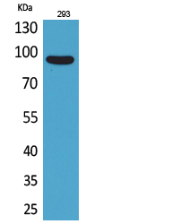
- 95kD
- Background:
- This gene encodes a transmembrane protein which contains three extracellular CUB domains and acts as a substrate for Src family kinases. The protein plays a role in the tyrosine phosphorylation-dependent regulation of cellular events that are involved in tumor invasion and metastasis. Alternative splicing results in multiple transcript variants of this gene. [provided by RefSeq, May 2013],
- Function:
- function:May be involved in cell adhesion and cell matrix association. May play a role in the regulation of anchorage versus migration or proliferation versus differentiation via its phosphorylation. May be a novel marker for leukemia diagnosis and for immature hematopoietic stem cell subsets. Belongs to the tetraspanin web involved in tumor progression and metastasis.,PTM:A soluble form may also be produced by proteolytic cleavage at the cell surface (shedding). Another peptide of 80 kDa (p80) is present in cultured keratinocytes probably due to tryptic cleavage at an unidentified site on its N-terminal side. Converted to p80 by plasmin, a trypsin-like protease.,PTM:N-glycosylated.,PTM:Phosphorylated on tyrosine by kinases of the SRC family such as SRC and YES as well as by the protein kinase C gamma/PRKCG. Dephosphorylated by phosphotyrosine phosphatases. Also phosphorylated by suramin
- Subcellular Location:
- [Isoform 1]: Cell membrane ; Single-pass membrane protein . Shedding may also lead to a soluble peptide.; [Isoform 3]: Secreted.
- Expression:
- Highly expressed in mitotic cells with low expression during interphase. Detected at highest levels in skeletal muscle and colon with lower levels in kidney, small intestine, placenta and lung. Up-regulated in a number of human tumor cell lines, as well as in colorectal cancer, breast carcinoma and lung cancer. Also expressed in cells with phenotypes reminiscent of mesenchymal stem cells and neural stem cells.
- June 19-2018
- WESTERN IMMUNOBLOTTING PROTOCOL
- June 19-2018
- IMMUNOHISTOCHEMISTRY-PARAFFIN PROTOCOL
- June 19-2018
- IMMUNOFLUORESCENCE PROTOCOL
- September 08-2020
- FLOW-CYTOMEYRT-PROTOCOL
- May 20-2022
- Cell-Based ELISA│解您多样本WB检测之困扰
- July 13-2018
- CELL-BASED-ELISA-PROTOCOL-FOR-ACETYL-PROTEIN
- July 13-2018
- CELL-BASED-ELISA-PROTOCOL-FOR-PHOSPHO-PROTEIN
- July 13-2018
- Antibody-FAQs
- Products Images

- Western Blot analysis of 293 cells using CDCP1 Polyclonal Antibody. Secondary antibody(catalog#:RS0002) was diluted at 1:20000

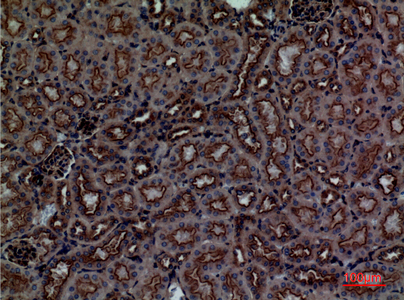
- Immunohistochemical analysis of paraffin-embedded mouse-kidney, antibody was diluted at 1:100

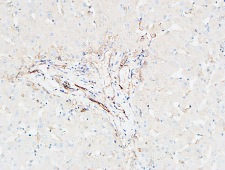
- Immunohistochemical analysis of paraffin-embedded Human Liver. 1, Antibody was diluted at 1:200(4° overnight). 2, High-pressure and temperature EDTA, pH8.0 was used for antigen retrieval. 3,Secondary antibody was diluted at 1:200(room temperature, 30min).

- Western blot analysis of lysate from 293 cells, using CDCP1 Antibody.


