Layilin Polyclonal Antibody
- Catalog No.:YT5222
- Applications:WB;IHC;IF;ELISA
- Reactivity:Human;Mouse;Rat
- Target:
- Layilin
- Gene Name:
- LAYN
- Protein Name:
- Layilin
- Human Gene Id:
- 143903
- Human Swiss Prot No:
- Q6UX15
- Mouse Gene Id:
- 244864
- Mouse Swiss Prot No:
- Q8C351
- Immunogen:
- The antiserum was produced against synthesized peptide derived from the Internal region of human LAYN. AA range:71-120
- Specificity:
- Layilin Polyclonal Antibody detects endogenous levels of Layilin protein.
- Formulation:
- Liquid in PBS containing 50% glycerol, 0.5% BSA and 0.02% sodium azide.
- Source:
- Polyclonal, Rabbit,IgG
- Dilution:
- WB 1:500 - 1:2000. IHC: 1:100-300 ELISA: 1:20000.. IF 1:50-200
- Purification:
- The antibody was affinity-purified from rabbit antiserum by affinity-chromatography using epitope-specific immunogen.
- Concentration:
- 1 mg/ml
- Storage Stability:
- -15°C to -25°C/1 year(Do not lower than -25°C)
- Other Name:
- LAYN;Layilin
- Observed Band(KD):
- 43kD
- Background:
- domain:The C-terminal domain interacts with the N-terminal domain of RDX.,function:Receptor for hyaluronate.,similarity:Contains 1 C-type lectin domain.,subcellular location:Colocalizes with TLN1 at the membrane ruffles.,subunit:Interacts with NF2, RDX and TLN1.,
- Function:
- domain:The C-terminal domain interacts with the N-terminal domain of RDX.,function:Receptor for hyaluronate.,similarity:Contains 1 C-type lectin domain.,subcellular location:Colocalizes with TLN1 at the membrane ruffles.,subunit:Interacts with NF2, RDX and TLN1.,
- Subcellular Location:
- Membrane ; Single-pass type I membrane protein . Colocalizes with TLN1 at the membrane ruffles. .
- Expression:
- Brain,
- June 19-2018
- WESTERN IMMUNOBLOTTING PROTOCOL
- June 19-2018
- IMMUNOHISTOCHEMISTRY-PARAFFIN PROTOCOL
- June 19-2018
- IMMUNOFLUORESCENCE PROTOCOL
- September 08-2020
- FLOW-CYTOMEYRT-PROTOCOL
- May 20-2022
- Cell-Based ELISA│解您多样本WB检测之困扰
- July 13-2018
- CELL-BASED-ELISA-PROTOCOL-FOR-ACETYL-PROTEIN
- July 13-2018
- CELL-BASED-ELISA-PROTOCOL-FOR-PHOSPHO-PROTEIN
- July 13-2018
- Antibody-FAQs
- Products Images
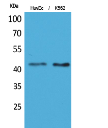
- Western Blot analysis of HuvEc, K562 cells using Layilin Polyclonal Antibody. Secondary antibody(catalog#:RS0002) was diluted at 1:20000
.jpg)
- Immunohistochemical analysis of paraffin-embedded rat-brain, antibody was diluted at 1:100
.jpg)
- Immunohistochemical analysis of paraffin-embedded rat-brain, antibody was diluted at 1:100
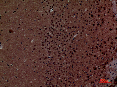
- Immunohistochemical analysis of paraffin-embedded mouse-brain, antibody was diluted at 1:100
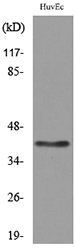
- Western blot analysis of lysate from HUVEC cells, using LAYN Antibody.


