Catalase Polyclonal Antibody
- Catalog No.:YT5155
- Applications:IF;WB;IHC;ELISA
- Reactivity:Human;Rat;Mouse;Fish
- Target:
- Catalase
- Fields:
- >>Tryptophan metabolism;>>Glyoxylate and dicarboxylate metabolism;>>Metabolic pathways;>>Carbon metabolism;>>FoxO signaling pathway;>>Peroxisome;>>Longevity regulating pathway;>>Longevity regulating pathway - multiple species;>>Amyotrophic lateral sclerosis;>>Pathways of neurodegeneration - multiple diseases;>>Chemical carcinogenesis - reactive oxygen species
- Gene Name:
- CAT
- Protein Name:
- Catalase
- Human Gene Id:
- 847
- Human Swiss Prot No:
- P04040
- Mouse Swiss Prot No:
- P24270
- Immunogen:
- The antiserum was produced against synthesized peptide derived from the C-terminal region of human CAT. AA range:478-527
- Specificity:
- Catalase Polyclonal Antibody detects endogenous levels of Catalase protein.
- Formulation:
- Liquid in PBS containing 50% glycerol, 0.5% BSA and 0.02% sodium azide.
- Source:
- Polyclonal, Rabbit,IgG
- Dilution:
- IF 1:50-200 WB 1:500 - 1:2000. IHC: 1:100-300 ELISA: 1:20000. Not yet tested in other applications.
- Purification:
- The antibody was affinity-purified from rabbit antiserum by affinity-chromatography using epitope-specific immunogen.
- Concentration:
- 1 mg/ml
- Storage Stability:
- -15°C to -25°C/1 year(Do not lower than -25°C)
- Other Name:
- CAT;Catalase
- Observed Band(KD):
- 60kD
- Background:
- This gene encodes catalase, a key antioxidant enzyme in the bodies defense against oxidative stress. Catalase is a heme enzyme that is present in the peroxisome of nearly all aerobic cells. Catalase converts the reactive oxygen species hydrogen peroxide to water and oxygen and thereby mitigates the toxic effects of hydrogen peroxide. Oxidative stress is hypothesized to play a role in the development of many chronic or late-onset diseases such as diabetes, asthma, Alzheimer's disease, systemic lupus erythematosus, rheumatoid arthritis, and cancers. Polymorphisms in this gene have been associated with decreases in catalase activity but, to date, acatalasemia is the only disease known to be caused by this gene. [provided by RefSeq, Oct 2009],
- Function:
- catalytic activity:2 H(2)O(2) = O(2) + 2 H(2)O.,cofactor:Heme group.,cofactor:NADP.,disease:Defects in CAT are the cause of acatalasia (ACATLAS) [MIM:115500]; also known as acatalasemia. This disease is characterized by absence of catalase activity in red cells and is often associated with ulcerating oral lesions.,function:Occurs in almost all aerobically respiring organisms and serves to protect cells from the toxic effects of hydrogen peroxide. Promotes growth of cells including T-cells, B-cells, myeloid leukemia cells, melanoma cells, mastocytoma cells and normal and transformed fibroblast cells.,online information:Catalase entry,PTM:The N-terminus is blocked.,similarity:Belongs to the catalase family.,subunit:Homotetramer.,
- Subcellular Location:
- Peroxisome.
- Expression:
- Brain,Cajal-Retzius cell,Erythrocyte,Eye,Fibroblast,Kidney,Liver,Placenta,Platelet,Skin,Uterus,
Chronic hypoxia and Cu2+ exposure induce gill remodeling of Largemouth bass through endoplasmic reticulum stress, mitochondrial damage and apoptosis AQUATIC TOXICOLOGY Qiao Liu, Hong Wang, Jiayu Ge, Lisen Li, Jie Luo, Kuo He, Haoxiao Yan, Xin Zhang, Rabia Tahir, Wei Luo, Shiyi Chen, Zhang Cheng, Liulan Zhao, Song Yang WB Fish Gill tissue
The protective effect of resveratrol on largemouth bass (Micropterus salmoides) during out-of-season spawning FISH & SHELLFISH IMMUNOLOGY Song Yang WB Fish ovarian tissue
- June 19-2018
- WESTERN IMMUNOBLOTTING PROTOCOL
- June 19-2018
- IMMUNOHISTOCHEMISTRY-PARAFFIN PROTOCOL
- June 19-2018
- IMMUNOFLUORESCENCE PROTOCOL
- September 08-2020
- FLOW-CYTOMEYRT-PROTOCOL
- May 20-2022
- Cell-Based ELISA│解您多样本WB检测之困扰
- July 13-2018
- CELL-BASED-ELISA-PROTOCOL-FOR-ACETYL-PROTEIN
- July 13-2018
- CELL-BASED-ELISA-PROTOCOL-FOR-PHOSPHO-PROTEIN
- July 13-2018
- Antibody-FAQs
- Products Images
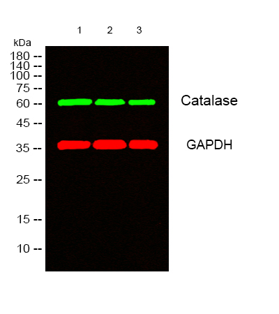
- Western blot analysis of lysates from 1) HT29, 2) A549, 3) HELA cells, (Green) primary antibody was diluted at 1:1000, 4°over night, secondary antibody(cat:RS23920)was diluted at 1:10000, 37° 1hour. (Red) GAPDH Monoclonal Antibody(2B8) (cat:YM3029) antibody was diluted at 1:5000 as loading control, 4° over night,secondary antibody(cat:RS23710)was diluted at 1:10000, 37° 1hour.
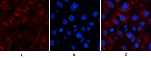
- Immunofluorescence analysis of human-liver tissue. 1,Catalase Polyclonal Antibody(red) was diluted at 1:200(4°C,overnight). 2, Cy3 labled Secondary antibody was diluted at 1:300(room temperature, 50min).3, Picture B: DAPI(blue) 10min. Picture A:Target. Picture B: DAPI. Picture C: merge of A+B
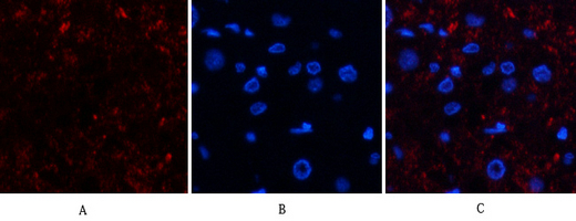
- Immunofluorescence analysis of human-kidney-cancer tissue. 1,Catalase Polyclonal Antibody(red) was diluted at 1:200(4°C,overnight). 2, Cy3 labled Secondary antibody was diluted at 1:300(room temperature, 50min).3, Picture B: DAPI(blue) 10min. Picture A:Target. Picture B: DAPI. Picture C: merge of A+B
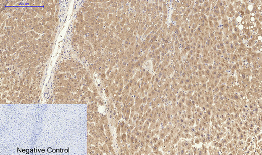
- Immunohistochemical analysis of paraffin-embedded Human-liver tissue. 1,Catalase Polyclonal Antibody was diluted at 1:200(4°C,overnight). 2, Sodium citrate pH 6.0 was used for antibody retrieval(>98°C,20min). 3,Secondary antibody was diluted at 1:200(room tempeRature, 30min). Negative control was used by secondary antibody only.
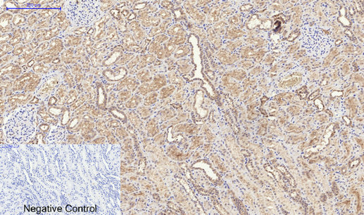
- Immunohistochemical analysis of paraffin-embedded Human-kidney tissue. 1,Catalase Polyclonal Antibody was diluted at 1:200(4°C,overnight). 2, Sodium citrate pH 6.0 was used for antibody retrieval(>98°C,20min). 3,Secondary antibody was diluted at 1:200(room tempeRature, 30min). Negative control was used by secondary antibody only.
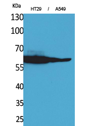
- Western Blot analysis of HT29, A549 cells using Catalase Polyclonal Antibody. Antibody was diluted at 1:1000. Secondary antibody(catalog#:RS0002) was diluted at 1:20000
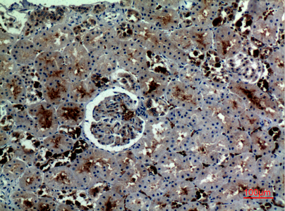
- Immunohistochemical analysis of paraffin-embedded human-kidney, antibody was diluted at 1:100


