IL-8 Polyclonal Antibody
- Catalog No.:YT5153
- Applications:IF;WB;IHC;ELISA
- Reactivity:Human
- Target:
- IL-8
- Fields:
- >>Cytokine-cytokine receptor interaction;>>Viral protein interaction with cytokine and cytokine receptor;>>Chemokine signaling pathway;>>NF-kappa B signaling pathway;>>Phospholipase D signaling pathway;>>Cellular senescence;>>Toll-like receptor signaling pathway;>>NOD-like receptor signaling pathway;>>RIG-I-like receptor signaling pathway;>>IL-17 signaling pathway;>>Non-alcoholic fatty liver disease;>>AGE-RAGE signaling pathway in diabetic complications;>>Alcoholic liver disease;>>Epithelial cell signaling in Helicobacter pylori infection;>>Pathogenic Escherichia coli infection;>>Shigellosis;>>Salmonella infection;>>Pertussis;>>Legionellosis;>>Yersinia infection;>>Chagas disease;>>Malaria;>>Amoebiasis;>>Hepatitis B;>>Human cytomegalovirus infection;>>Influenza A;>>Kaposi sarcoma-associated herpesvirus infection;>>Coronavirus disease - COVID-19;>>Pathways in cancer;>>Transcriptional misregulation in cancer;>>Bladder cancer;>>Rheumatoid arthritis;>>Lipid and atherosclerosis
- Gene Name:
- IL8 CXCL8
- Protein Name:
- Interleukin-8
- Human Gene Id:
- 3576
- Human Swiss Prot No:
- P10145
- Immunogen:
- The antiserum was produced against synthesized peptide derived from the C-terminal region of human IL8. AA range:50-99
- Specificity:
- IL-8 Polyclonal Antibody detects endogenous levels of IL-8 protein.
- Formulation:
- Liquid in PBS containing 50% glycerol, 0.5% BSA and 0.02% sodium azide.
- Source:
- Polyclonal, Rabbit,IgG
- Dilution:
- IF 1:50-200 WB 1:500 - 1:2000. IHC: 1:100-300 ELISA: 1:20000. Not yet tested in other applications.
- Purification:
- The antibody was affinity-purified from rabbit antiserum by affinity-chromatography using epitope-specific immunogen.
- Concentration:
- 1 mg/ml
- Storage Stability:
- -15°C to -25°C/1 year(Do not lower than -25°C)
- Other Name:
- IL8;CXCL8;Interleukin-8;IL-8;C-X-C motif chemokine 8;Emoctakin;Granulocyte chemotactic protein 1;GCP-1;Monocyte-derived neutrophil chemotactic factor;MDNCF;Monocyte-derived neutrophil-activating peptide;MONAP;Neutrophil-activating protein 1;NAP-1;Protein 3-10C;T-cell chemotactic factor
- Observed Band(KD):
- 11kD
- Background:
- The protein encoded by this gene is a member of the CXC chemokine family. This chemokine is one of the major mediators of the inflammatory response. This chemokine is secreted by several cell types. It functions as a chemoattractant, and is also a potent angiogenic factor. This gene is believed to play a role in the pathogenesis of bronchiolitis, a common respiratory tract disease caused by viral infection. This gene and other ten members of the CXC chemokine gene family form a chemokine gene cluster in a region mapped to chromosome 4q. [provided by RefSeq, Jul 2008],
- Function:
- function:IL-8 is a chemotactic factor that attracts neutrophils, basophils, and T-cells, but not monocytes. It is also involved in neutrophil activation. It is released from several cell types in response to an inflammatory stimulus. IL-8(6-77) has a 5-10-fold higher activity on neutrophil activation, IL-8(5-77) has increased activity on neutrophil activation and IL-8(7-77) has a higher affinity to receptors CXCR1 and CXCR2 as compared to IL-8(1-77), respectively.,online information:Interleukin-8 entry,PTM:Several N-terminal processed forms are produced by proteolytic cleavage after secretion from at least peripheral blood monocytes, leukcocytes and endothelial cells. In general, IL-8(1-77) is referred to as interleukin-8. IL-8(6-77) is the most promiment form.,similarity:Belongs to the intercrine alpha (chemokine CxC) family.,subunit:Homodimer.,
- Subcellular Location:
- Secreted.
- Expression:
- Chronic myeloid leukemia cell,Kidney,Lung,Lung carcinoma,Neutrophil,Periphe
Oridonin Delays Aging Through the AKT Signaling Pathway WB Human /WI-38 cell
Targeting myeloid derived suppressor cells reverts immune suppression and sensitizes BRAF-mutant papillary thyroid cancer to MAPK inhibitors Nat Commun. 2022 Mar;13(1):1-18. IF,IHC Mouse 1:500 K1 cell-xenograft
Human Novel MicroRNA Seq-915_x4024 in Keratinocytes Contributes to Skin Regeneration by Suppressing Scar Formation. Molecular Therapy-Nucleic Acids Mol Ther-Nucl Acids. 2019 Mar;14:410 WB Human HaCaT cell
Association between higher expression of interleukin-8 (IL-8) and haplotype −353A/−251A/+678T of IL-8 gene with preeclampsia: A case–control study. MEDICINE Medicine. 2016 Dec; 95(52): e5537 IHC Human 1:200 Placental tissue
CXCL5 as an autocrine or paracrine cytokine is associated with proliferation and migration of hepatoblastoma HepG2 cells. Oncology Letters Oncol Lett. 2017 Dec;14(6):7977-7985 WB Human 1:1000 HepG2 cells
Hui, Zhi, and Zhe Li. "Continuous veno-venous hemofiltration for septic shock." INTERNATIONAL JOURNAL OF CLINICAL AND EXPERIMENTAL MEDICINE 10.10 (2017): 14983-14988.
Li, Na, et al. "Asparaginyl endopeptidase may promote liver sinusoidal endothelial cell angiogenesis via PI3K/Akt pathway." Revista espanola de enfermedades digestivas: organo oficial de la Sociedad Espanola de Patologia Digestiva111 (2018).
Helicobacter pylori with trx1 high expression promotes gastric diseases via upregulating the IL23A/NF-κB/IL8 pathway HELICOBACTER Xin Guan IHC Human 1:200 antrum tissue
- June 19-2018
- WESTERN IMMUNOBLOTTING PROTOCOL
- June 19-2018
- IMMUNOHISTOCHEMISTRY-PARAFFIN PROTOCOL
- June 19-2018
- IMMUNOFLUORESCENCE PROTOCOL
- September 08-2020
- FLOW-CYTOMEYRT-PROTOCOL
- May 20-2022
- Cell-Based ELISA│解您多样本WB检测之困扰
- July 13-2018
- CELL-BASED-ELISA-PROTOCOL-FOR-ACETYL-PROTEIN
- July 13-2018
- CELL-BASED-ELISA-PROTOCOL-FOR-PHOSPHO-PROTEIN
- July 13-2018
- Antibody-FAQs
- Products Images

- Immunofluorescence analysis of Hela cell. 1,IL-8 Polyclonal Antibody(green) was diluted at 1:200(4° overnight). 2, Goat Anti Rabbit Alexa Fluor 488 Catalog:RS3211 was diluted at 1:1000(room temperature, 50min). 3 DAPI(blue) 10min.
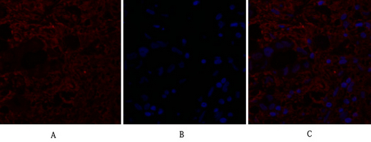
- Immunofluorescence analysis of human-breast-cancer tissue. 1,IL-8 Polyclonal Antibody(red) was diluted at 1:200(4°C,overnight). 2, Cy3 labled Secondary antibody was diluted at 1:300(room temperature, 50min).3, Picture B: DAPI(blue) 10min. Picture A:Target. Picture B: DAPI. Picture C: merge of A+B
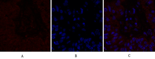
- Immunofluorescence analysis of human-liver-cancer tissue. 1,IL-8 Polyclonal Antibody(red) was diluted at 1:200(4°C,overnight). 2, Cy3 labled Secondary antibody was diluted at 1:300(room temperature, 50min).3, Picture B: DAPI(blue) 10min. Picture A:Target. Picture B: DAPI. Picture C: merge of A+B
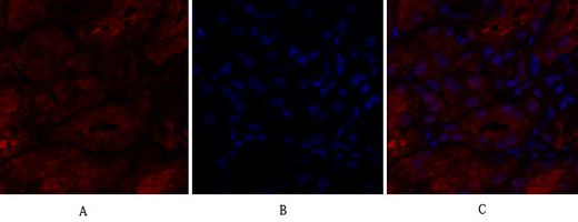
- Immunofluorescence analysis of human-kidney tissue. 1,IL-8 Polyclonal Antibody(red) was diluted at 1:200(4°C,overnight). 2, Cy3 labled Secondary antibody was diluted at 1:300(room temperature, 50min).3, Picture B: DAPI(blue) 10min. Picture A:Target. Picture B: DAPI. Picture C: merge of A+B
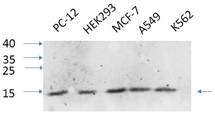
- Western Blot analysis of various cells using primary antibody diluted at 1:1000(4°C overnight). Secondary antibody:Goat Anti-rabbit IgG IRDye 800( diluted at 1:5000, 25°C, 1 hour). Cell lysate was extracted by Minute™ Plasma Membrane Protein Isolation and Cell Fractionation Kit(SM-005, Inventbiotech,MN,USA).
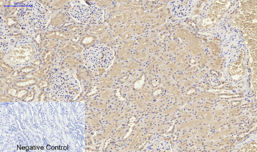
- Immunohistochemical analysis of paraffin-embedded Human-kidney tissue. 1,IL-8 Polyclonal Antibody was diluted at 1:200(4°C,overnight). 2, Sodium citrate pH 6.0 was used for antibody retrieval(>98°C,20min). 3,Secondary antibody was diluted at 1:200(room tempeRature, 30min). Negative control was used by secondary antibody only.
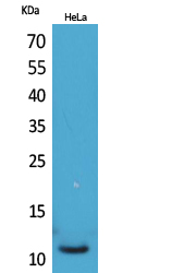
- Western Blot analysis of HeLa cells using IL-8 Polyclonal Antibody. Secondary antibody(catalog#:RS0002) was diluted at 1:20000
.jpg)
- Immunohistochemical analysis of paraffin-embedded human-skin, antibody was diluted at 1:100
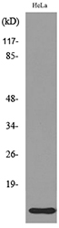
- Western blot analysis of lysate from HeLa cells, using IL8 Antibody.


