WIP Polyclonal Antibody
- Catalog No.:YT4905
- Applications:WB;IHC;IF;ELISA
- Reactivity:Human;Mouse;Rat
- Target:
- WIP
- Fields:
- >>Endocytosis;>>Pathogenic Escherichia coli infection;>>Yersinia infection
- Gene Name:
- WIPF1
- Protein Name:
- WAS/WASL-interacting protein family member 1
- Human Gene Id:
- 7456
- Human Swiss Prot No:
- O43516
- Mouse Gene Id:
- 215280
- Mouse Swiss Prot No:
- Q8K1I7
- Rat Gene Id:
- 117538
- Rat Swiss Prot No:
- Q6IN36
- Immunogen:
- The antiserum was produced against synthesized peptide derived from human WIPF1. AA range:421-470
- Specificity:
- WIP Polyclonal Antibody detects endogenous levels of WIP protein.
- Formulation:
- Liquid in PBS containing 50% glycerol, 0.5% BSA and 0.02% sodium azide.
- Source:
- Polyclonal, Rabbit,IgG
- Dilution:
- WB 1:500 - 1:2000. IHC 1:100 - 1:300. ELISA: 1:5000.. IF 1:50-200
- Purification:
- The antibody was affinity-purified from rabbit antiserum by affinity-chromatography using epitope-specific immunogen.
- Concentration:
- 1 mg/ml
- Storage Stability:
- -15°C to -25°C/1 year(Do not lower than -25°C)
- Other Name:
- WIPF1;WASPIP;WIP;WAS/WASL-interacting protein family member 1;Protein PRPL-2;Wiskott-Aldrich syndrome protein-interacting protein;WASP-interacting protein
- Observed Band(KD):
- 52kD
- Background:
- This gene encodes a protein that plays an important role in the organization of the actin cytoskeleton. The encoded protein binds to a region of Wiskott-Aldrich syndrome protein that is frequently mutated in Wiskott-Aldrich syndrome, an X-linked recessive disorder. Impairment of the interaction between these two proteins may contribute to the disease. Two transcript variants encoding the same protein have been identified for this gene. [provided by RefSeq, Jul 2008],
- Function:
- domain:Binds to WAS within the N-terminal region 170, at a site distinct from the CDC42-binding site.,function:May have direct activity on the actin cytoskeleton. Induces actin polymerization and redistribution. Contributes with NCK1 and GRB2 in the recruitment and activation of WASL. May participate in regulating the subcellular localization of WASL, resulting in the disassembly of stress fibers in favor of filopodia formation (By similarity). Plays an important role in the intracellular motility of vaccinia virus by functioning as an adapter for recruiting WASL to vaccinia virus.,miscellaneous:Recruited to PIP5K-induced vesicle surfaces in the absence of functional WASL.,similarity:Belongs to the verprolin family.,similarity:Contains 1 WH2 domain.,subcellular location:Vesicle surfaces and along actin tails. Co-localized with actin stress fibers. When co-expressed with WASL, no longer a
- Subcellular Location:
- Cytoplasmic vesicle . Cytoplasm, cytoskeleton . Cell projection, ruffle . Vesicle surfaces and along actin tails. Colocalizes with actin stress fibers. When coexpressed with WASL, no longer associated with actin filaments but accumulated in perinuclear and cortical areas like WASL (By similarity). .
- Expression:
- Highly expressed in peripheral blood mononuclear cells, spleen, placenta, small intestine, colon and thymus. Lower expression in ovary, heart, brain, lung, liver, skeletal muscle, kidney, pancreas, prostate and testis.
- June 19-2018
- WESTERN IMMUNOBLOTTING PROTOCOL
- June 19-2018
- IMMUNOHISTOCHEMISTRY-PARAFFIN PROTOCOL
- June 19-2018
- IMMUNOFLUORESCENCE PROTOCOL
- September 08-2020
- FLOW-CYTOMEYRT-PROTOCOL
- May 20-2022
- Cell-Based ELISA│解您多样本WB检测之困扰
- July 13-2018
- CELL-BASED-ELISA-PROTOCOL-FOR-ACETYL-PROTEIN
- July 13-2018
- CELL-BASED-ELISA-PROTOCOL-FOR-PHOSPHO-PROTEIN
- July 13-2018
- Antibody-FAQs
- Products Images
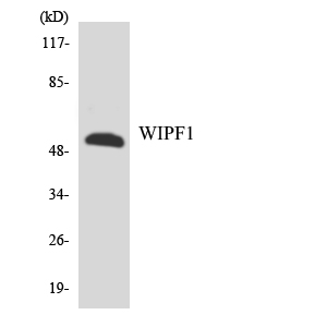
- Western blot analysis of the lysates from HT-29 cells using WIPF1 antibody.

- Immunohistochemical analysis of paraffin-embedded human Gastric adenocarcinoma. 1, Antibody was diluted at 1:200(4° overnight). 2, Tris-EDTA,pH9.0 was used for antigen retrieval. 3,Secondary antibody was diluted at 1:200(room temperature, 45min).


