SOCS-1 Polyclonal Antibody
- Catalog No.:YT4362
- Applications:IF;WB;IHC;ELISA
- Reactivity:Human;Mouse;Rat
- Target:
- SOCS-1
- Fields:
- >>Ubiquitin mediated proteolysis;>>Osteoclast differentiation;>>JAK-STAT signaling pathway;>>Insulin signaling pathway;>>Prolactin signaling pathway;>>Type II diabetes mellitus;>>Growth hormone synthesis, secretion and action;>>Toxoplasmosis;>>MicroRNAs in cancer
- Gene Name:
- SOCS1
- Protein Name:
- Suppressor of cytokine signaling 1
- Human Gene Id:
- 8651
- Human Swiss Prot No:
- O15524
- Mouse Gene Id:
- 12703
- Mouse Swiss Prot No:
- O35716
- Rat Gene Id:
- 252971
- Rat Swiss Prot No:
- Q9QX78
- Immunogen:
- The antiserum was produced against synthesized peptide derived from human SOCS-1. AA range:49-98
- Specificity:
- SOCS-1 Polyclonal Antibody detects endogenous levels of SOCS-1 protein.
- Formulation:
- Liquid in PBS containing 50% glycerol, 0.5% BSA and 0.02% sodium azide.
- Source:
- Polyclonal, Rabbit,IgG
- Dilution:
- IF 1:50-200 WB 1:500-2000, ELISA 1:10000-20000 IHC 1:50-300
- Purification:
- The antibody was affinity-purified from rabbit antiserum by affinity-chromatography using epitope-specific immunogen.
- Concentration:
- 1 mg/ml
- Storage Stability:
- -15°C to -25°C/1 year(Do not lower than -25°C)
- Other Name:
- SOCS1;SSI1;TIP3;Suppressor of cytokine signaling 1;SOCS-1;JAK-binding protein;JAB;STAT-induced STAT inhibitor 1;SSI-1;Tec-interacting protein 3;TIP-3
- Observed Band(KD):
- 38kD
- Background:
- This gene encodes a member of the STAT-induced STAT inhibitor (SSI), also known as suppressor of cytokine signaling (SOCS), family. SSI family members are cytokine-inducible negative regulators of cytokine signaling. The expression of this gene can be induced by a subset of cytokines, including IL2, IL3 erythropoietin (EPO), CSF2/GM-CSF, and interferon (IFN)-gamma. The protein encoded by this gene functions downstream of cytokine receptors, and takes part in a negative feedback loop to attenuate cytokine signaling. Knockout studies in mice suggested the role of this gene as a modulator of IFN-gamma action, which is required for normal postnatal growth and survival. [provided by RefSeq, Jul 2008],
- Function:
- domain:The ESS and SH2 domains are required for JAK phosphotyrosine binding. Further interaction with the KIR domain is necessary for signal and kinase inhibition.,domain:The SOCS box domain mediates the interaction with the Elongin BC complex, an adapter module in different E3 ubiquitin ligase complexes. The Elongin BC complex binding domain is also known as BC-box with the consensus [APST]-L-x(3)-C-x(3)-[AILV] and is part of the SOCS box.,function:SOCS family proteins form part of a classical negative feedback system that regulates cytokine signal transduction. SOCS1 is involved in negative regulation of cytokines that signal through the JAK/STAT3 pathway. Through binding to JAKs, inhibits their kinase activity. In vitro, also suppresses Tec protein-tyrosine activity. Appears to be a major regulator of signaling by interleukin 6 (IL6) and leukemia inhibitory factor (LIF). Regulates int
- Subcellular Location:
- Nucleus . Cytoplasmic vesicle . Detected in perinuclear cytoplasmic vesicles upon interaction with FGFR3.
- Expression:
- Expressed in all tissues with high expression in spleen, small intestine and peripheral blood leukocytes.
Targeting androgen receptor with ASC-J9 attenuates cardiac injury and dysfunction in experimental autoimmune myocarditis by reducing M1-like macrophage. BIOCHEMICAL AND BIOPHYSICAL RESEARCH COMMUNICATIONS Biochem Bioph Res Co. 2017 Apr;485:746 WB Mouse 1:500 Raw264.7 macrophages
Danzhi Jiangtang Capsule ameliorates kidney injury via inhibition of the JAK-STAT signaling pathway and increased antioxidant capacity in STZ-induced diabetic nephropathy rats. BioScience Trends 2018 Dec 28 WB Rat 1:300 Renal tissues
DJC Suppresses Advanced Glycation End Products-Induced JAK-STAT Signaling and ROS in Mesangial Cells. Evidence-based Complementary and Alternative Medicine Evid-Based Compl Alt. 2017;2017:2942830 WB Mouse glomerular mesangial cells (GMCs)
Targeting androgen receptor with ASC-J9 attenuates cardiac injury and dysfunction in experimental autoimmune myocarditis by reducing M1-like macrophage. BIOCHEMICAL AND BIOPHYSICAL RESEARCH COMMUNICATIONS Biochem Bioph Res Co. 2017 Apr;485:746 WB Mouse 1:500 Raw264.7 macrophages
miRNA-331-3p Affects the Proliferation, Metastasis, and Invasion of Osteosarcoma through SOCS1/JAK2/STAT3 Journal of Oncology Bing Wu WB, IHC Mouse, Human
- June 19-2018
- WESTERN IMMUNOBLOTTING PROTOCOL
- June 19-2018
- IMMUNOHISTOCHEMISTRY-PARAFFIN PROTOCOL
- June 19-2018
- IMMUNOFLUORESCENCE PROTOCOL
- September 08-2020
- FLOW-CYTOMEYRT-PROTOCOL
- May 20-2022
- Cell-Based ELISA│解您多样本WB检测之困扰
- July 13-2018
- CELL-BASED-ELISA-PROTOCOL-FOR-ACETYL-PROTEIN
- July 13-2018
- CELL-BASED-ELISA-PROTOCOL-FOR-PHOSPHO-PROTEIN
- July 13-2018
- Antibody-FAQs
- Products Images
- Sun, Min, et al. "DJC Suppresses Advanced Glycation End Products-Induced JAK-STAT Signaling and ROS in Mesangial Cells." Evidence-Based Complementary and Alternative Medicine 2017 (2017).
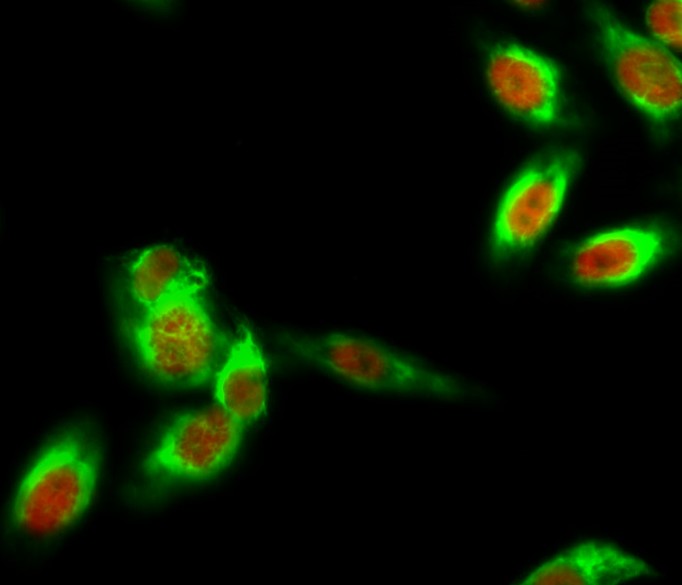
- Immunofluorescence analysis of Hela cell. 1,SOCS-1 Polyclonal Antibody(red) was diluted at 1:200(4° overnight). β-Tubulin Monoclonal Antibody(5G3)(green) was diluted at 1:200(4° overnight). 2, Goat Anti Rabbit Alexa Fluor 594 Catalog:RS3611 was diluted at 1:1000(room temperature, 50min). Goat Anti Mouse Alexa Fluor 488 Catalog:RS3208 was diluted at 1:1000(room temperature, 50min).
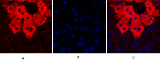
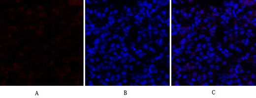
- Immunofluorescence analysis of mouse-liver tissue. 1,SOCS-1 Polyclonal Antibody(red) was diluted at 1:200(4°C,overnight). 2, Cy3 labled Secondary antibody was diluted at 1:300(room temperature, 50min).3, Picture B: DAPI(blue) 10min. Picture A:Target. Picture B: DAPI. Picture C: merge of A+B
- Immunofluorescence analysis of mouse-liver tissue. 1,SOCS-1 Polyclonal Antibody(red) was diluted at 1:200(4°C,overnight). 2, Cy3 labled Secondary antibody was diluted at 1:300(room temperature, 50min).3, Picture B: DAPI(blue) 10min. Picture A:Target. Picture B: DAPI. Picture C: merge of A+B
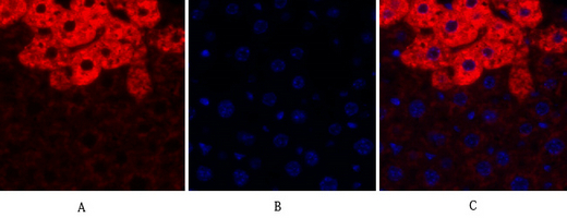
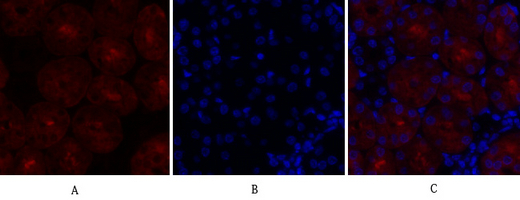
- Immunofluorescence analysis of mouse-lung tissue. 1,SOCS-1 Polyclonal Antibody(red) was diluted at 1:200(4°C,overnight). 2, Cy3 labled Secondary antibody was diluted at 1:300(room temperature, 50min).3, Picture B: DAPI(blue) 10min. Picture A:Target. Picture B: DAPI. Picture C: merge of A+B
- Immunofluorescence analysis of mouse-kidney tissue. 1,SOCS-1 Polyclonal Antibody(red) was diluted at 1:200(4°C,overnight). 2, Cy3 labled Secondary antibody was diluted at 1:300(room temperature, 50min).3, Picture B: DAPI(blue) 10min. Picture A:Target. Picture B: DAPI. Picture C: merge of A+B
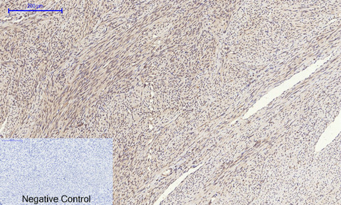
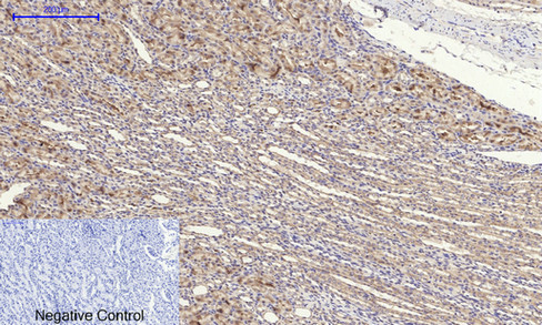
- Immunohistochemical analysis of paraffin-embedded Human-uterus tissue. 1,SOCS-1 Polyclonal Antibody was diluted at 1:200(4°C,overnight). 2, Sodium citrate pH 6.0 was used for antibody retrieval(>98°C,20min). 3,Secondary antibody was diluted at 1:200(room tempeRature, 30min). Negative control was used by secondary antibody only.

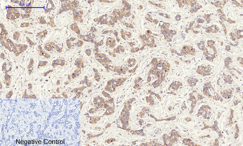
- Immunohistochemical analysis of paraffin-embedded Human-uterus-cancer tissue. 1,SOCS-1 Polyclonal Antibody was diluted at 1:200(4°C,overnight). 2, Sodium citrate pH 6.0 was used for antibody retrieval(>98°C,20min). 3,Secondary antibody was diluted at 1:200(room tempeRature, 30min). Negative control was used by secondary antibody only.
- Immunohistochemical analysis of paraffin-embedded Human-liver-cancer tissue. 1,SOCS-1 Polyclonal Antibody was diluted at 1:200(4°C,overnight). 2, Sodium citrate pH 6.0 was used for antibody retrieval(>98°C,20min). 3,Secondary antibody was diluted at 1:200(room tempeRature, 30min). Negative control was used by secondary antibody only.
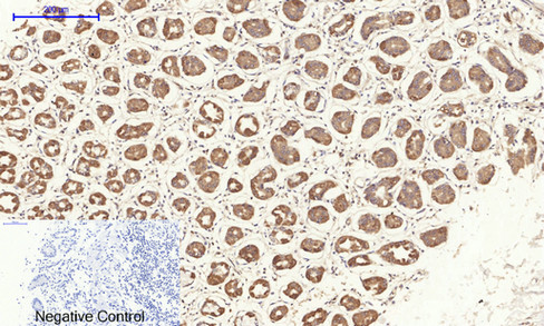
- Immunohistochemical analysis of paraffin-embedded Human-stomach tissue. 1,SOCS-1 Polyclonal Antibody was diluted at 1:200(4°C,overnight). 2, Sodium citrate pH 6.0 was used for antibody retrieval(>98°C,20min). 3,Secondary antibody was diluted at 1:200(room tempeRature, 30min). Negative control was used by secondary antibody only.
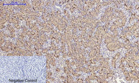
- Immunohistochemical analysis of paraffin-embedded Rat-kidney tissue. 1,SOCS-1 Polyclonal Antibody was diluted at 1:200(4°C,overnight). 2, Sodium citrate pH 6.0 was used for antibody retrieval(>98°C,20min). 3,Secondary antibody was diluted at 1:200(room tempeRature, 30min). Negative control was used by secondary antibody only.
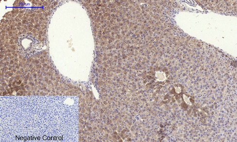
- Immunohistochemical analysis of paraffin-embedded Mouse-liver tissue. 1,SOCS-1 Polyclonal Antibody was diluted at 1:200(4°C,overnight). 2, Sodium citrate pH 6.0 was used for antibody retrieval(>98°C,20min). 3,Secondary antibody was diluted at 1:200(room tempeRature, 30min). Negative control was used by secondary antibody only.
- Immunohistochemical analysis of paraffin-embedded Mouse-kidney tissue. 1,SOCS-1 Polyclonal Antibody was diluted at 1:200(4°C,overnight). 2, Sodium citrate pH 6.0 was used for antibody retrieval(>98°C,20min). 3,Secondary antibody was diluted at 1:200(room tempeRature, 30min). Negative control was used by secondary antibody only.
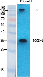
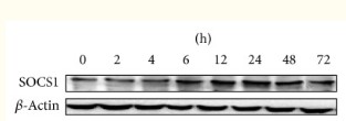
- Western Blot analysis of various cells using SOCS-1 Polyclonal Antibody diluted at 1:2000
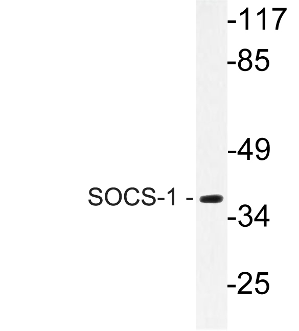
- Western blot analysis of lysate from Jurkat cells, using SOCS-1 antibody.


