SEMA3F Polyclonal Antibody
- Catalog No.:YT4234
- Applications:WB;ELISA;IHC
- Reactivity:Human;Mouse;Rat
- Target:
- SEMA3F
- Fields:
- >>Axon guidance
- Gene Name:
- SEMA3F
- Protein Name:
- Semaphorin-3F
- Human Gene Id:
- 6405
- Human Swiss Prot No:
- Q13275
- Mouse Gene Id:
- 20350
- Mouse Swiss Prot No:
- O88632
- Immunogen:
- The antiserum was produced against synthesized peptide derived from human SEMA3F. AA range:734-783
- Specificity:
- SEMA3F Polyclonal Antibody detects endogenous levels of SEMA3F protein.
- Formulation:
- Liquid in PBS containing 50% glycerol, 0.5% BSA and 0.02% sodium azide.
- Source:
- Polyclonal, Rabbit,IgG
- Dilution:
- WB 1:500-2000;IHC 1:50-300; ELISA 2000-20000
- Purification:
- The antibody was affinity-purified from rabbit antiserum by affinity-chromatography using epitope-specific immunogen.
- Concentration:
- 1 mg/ml
- Storage Stability:
- -15°C to -25°C/1 year(Do not lower than -25°C)
- Other Name:
- SEMA3F;Semaphorin-3F;Sema III/F;Semaphorin IV;Sema IV
- Observed Band(KD):
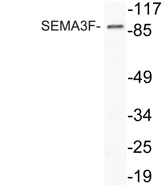
- 88kD
- Background:
- This gene encodes a member of the semaphorin III family of secreted signaling proteins that are involved in axon guidance during neuronal development. The encoded protein contains an N-terminal Sema domain, an immunoglobulin loop and a C-terminal basic domain. This gene is expressed by the endothelial cells where it was found to act in an autocrine fashion to induce apoptosis, inhibit cell proliferation and survival, and function as an anti-tumorigenic agent. Alternative splicing results in multiple transcript variants encoding different isoforms. [provided by RefSeq, Jan 2016],
- Function:
- developmental stage:Detected as early as embryonic day 10.,function:May play a role in cell motility and cell adhesion.,similarity:Belongs to the semaphorin family.,similarity:Contains 1 Ig-like C2-type (immunoglobulin-like) domain.,similarity:Contains 1 Sema domain.,tissue specificity:Expressed abundantly but differentially in a variety of neural and nonneural tissues. There is high expression in mammary gland, kidney, fetal brain, and lung and lower expression in heart and liver.,
- Subcellular Location:
- Secreted .
- Expression:
- Expressed abundantly but differentially in a variety of neural and nonneural tissues. There is high expression in mammary gland, kidney, fetal brain, and lung and lower expression in heart and liver.
- June 19-2018
- WESTERN IMMUNOBLOTTING PROTOCOL
- June 19-2018
- IMMUNOHISTOCHEMISTRY-PARAFFIN PROTOCOL
- June 19-2018
- IMMUNOFLUORESCENCE PROTOCOL
- September 08-2020
- FLOW-CYTOMEYRT-PROTOCOL
- May 20-2022
- Cell-Based ELISA│解您多样本WB检测之困扰
- July 13-2018
- CELL-BASED-ELISA-PROTOCOL-FOR-ACETYL-PROTEIN
- July 13-2018
- CELL-BASED-ELISA-PROTOCOL-FOR-PHOSPHO-PROTEIN
- July 13-2018
- Antibody-FAQs
- Products Images

- Western blot analysis of lysate from HeLa cells, using SEMA3F antibody.

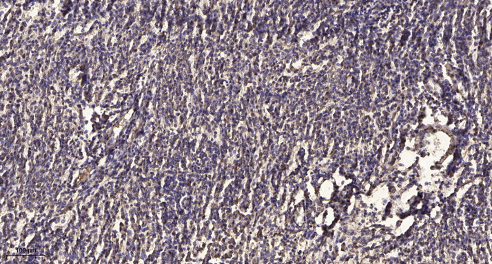
- Immunohistochemical analysis of paraffin-embedded human meningioma. 1, Antibody was diluted at 1:200(4° overnight). 2, Tris-EDTA,pH9.0 was used for antigen retrieval. 3,Secondary antibody was diluted at 1:200(room temperature, 45min).


