Saposin Polyclonal Antibody
- Catalog No.:YT4213
- Applications:WB;IHC;IF;ELISA
- Reactivity:Human;Rat;Mouse;
- Target:
- Saposin
- Fields:
- >>Sphingolipid metabolism;>>Lysosome
- Gene Name:
- PSAP
- Protein Name:
- Proactivator polypeptide
- Human Gene Id:
- 5660
- Human Swiss Prot No:
- P07602
- Mouse Swiss Prot No:
- Q61207
- Immunogen:
- The antiserum was produced against synthesized peptide derived from human PSAP. AA range:307-356
- Specificity:
- Saposin Polyclonal Antibody detects endogenous levels of Saposin protein.
- Formulation:
- Liquid in PBS containing 50% glycerol, 0.5% BSA and 0.02% sodium azide.
- Source:
- Polyclonal, Rabbit,IgG
- Dilution:
- WB 1:500 - 1:2000. IHC 1:100 - 1:300. ELISA: 1:20000.. IF 1:50-200
- Purification:
- The antibody was affinity-purified from rabbit antiserum by affinity-chromatography using epitope-specific immunogen.
- Concentration:
- 1 mg/ml
- Storage Stability:
- -15°C to -25°C/1 year(Do not lower than -25°C)
- Other Name:
- PSAP;GLBA;SAP1;Proactivator polypeptide
- Observed Band(KD):
- 58kD
- Background:
- This gene encodes a highly conserved preproprotein that is proteolytically processed to generate four main cleavage products including saposins A, B, C, and D. Each domain of the precursor protein is approximately 80 amino acid residues long with nearly identical placement of cysteine residues and glycosylation sites. Saposins A-D localize primarily to the lysosomal compartment where they facilitate the catabolism of glycosphingolipids with short oligosaccharide groups. The precursor protein exists both as a secretory protein and as an integral membrane protein and has neurotrophic activities. Mutations in this gene have been associated with Gaucher disease and metachromatic leukodystrophy. Alternative splicing results in multiple transcript variants, at least one of which encodes an isoform that is proteolytically processed. [provided by RefSeq, Feb 2016],
- Function:
- alternative products:Additional isoforms seem to exist,disease:Defects in PSAP are the cause of combined saposin deficiency (CSAPD) [MIM:611721]; also known as prosaposin deficiency. CSAPD is due to absence of all saposins, leading to a fatal storage disorder with hepatosplenomegaly and severe neurological involvement.,disease:Defects in PSAP saposin-A region are the cause of atypical Krabbe disease (AKRD) [MIM:611722]. AKRD is a disorder of galactosylceramide metabolism. AKRD features include progressive encephalopathy and abnormal myelination in the cerebral white matter resembling Krabbe disease.,disease:Defects in PSAP saposin-B region are the cause of a variant of metachromatic leukodystrophy (MLD) [MIM:249900].,disease:Defects in PSAP saposin-C region are the cause of atypical Gaucher disease (AGD) [MIM:610539]. Affected individuals have marked glucosylceramide accumulation in the
- Subcellular Location:
- Lysosome .; [Prosaposin]: Secreted . Secreted as a fully glycosylated 70 kDa protein composed of complex glycans. .
- Expression:
- Brain,Eye,Kidney,Liver,Milk,Peripheral Nervous System,Skin,Synovial membrane,Urine,
- June 19-2018
- WESTERN IMMUNOBLOTTING PROTOCOL
- June 19-2018
- IMMUNOHISTOCHEMISTRY-PARAFFIN PROTOCOL
- June 19-2018
- IMMUNOFLUORESCENCE PROTOCOL
- September 08-2020
- FLOW-CYTOMEYRT-PROTOCOL
- May 20-2022
- Cell-Based ELISA│解您多样本WB检测之困扰
- July 13-2018
- CELL-BASED-ELISA-PROTOCOL-FOR-ACETYL-PROTEIN
- July 13-2018
- CELL-BASED-ELISA-PROTOCOL-FOR-PHOSPHO-PROTEIN
- July 13-2018
- Antibody-FAQs
- Products Images
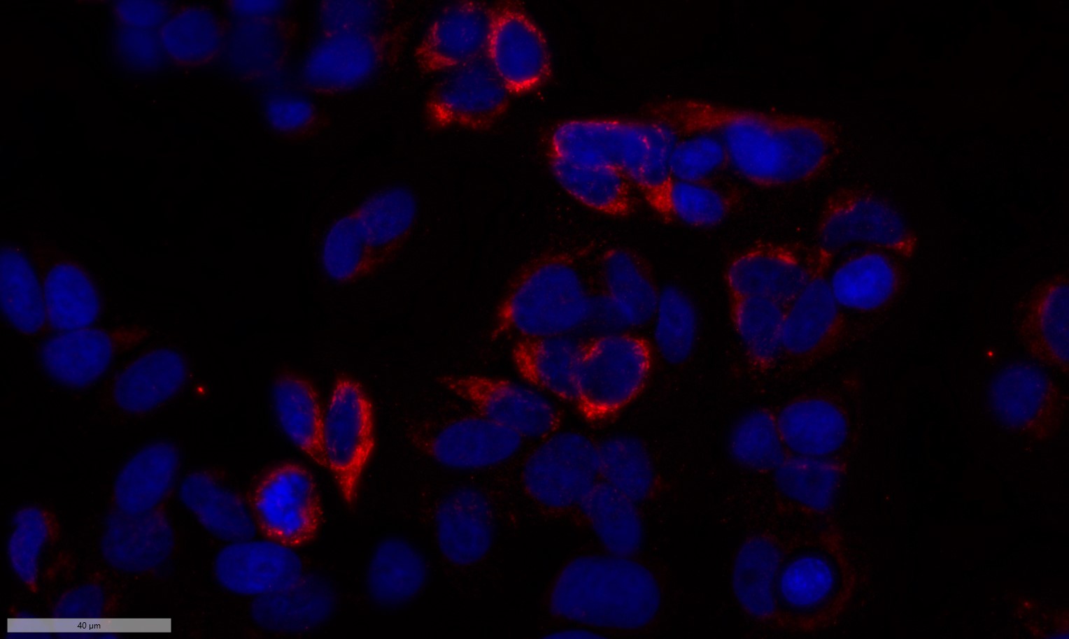
- Immunofluorescence analysis of MCF7 cell. 1,primary Antibody was diluted at 1:100(4°C overnight). 2, Goat Anti Rabbit IgG (H&L) - AFluor 594 Secondary antibody(catalog No: RS3611) was diluted at 1:500(room temperature, 50min).
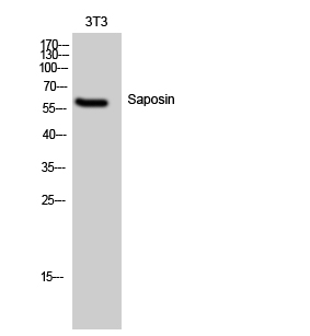
- Western Blot analysis of 3T3 cells using Saposin Polyclonal Antibody
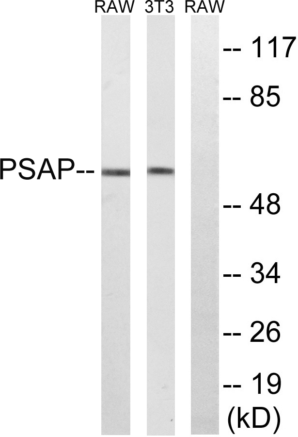
- Western blot analysis of lysates from NIH/3T3 and RAW264.7 cells, using PSAP Antibody. The lane on the right is blocked with the synthesized peptide.
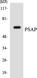
- Western blot analysis of the lysates from HeLa cells using PSAP antibody.
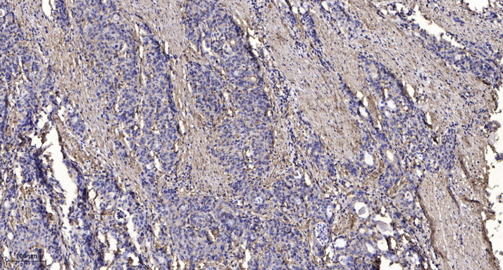
- Immunohistochemical analysis of paraffin-embedded human Gastric adenocarcinoma. 1, Antibody was diluted at 1:200(4° overnight). 2, Tris-EDTA,pH9.0 was used for antigen retrieval. 3,Secondary antibody was diluted at 1:200(room temperature, 45min).


