RXRγ Polyclonal Antibody
- Catalog No.:YT4195
- Applications:WB;IHC;IF;ELISA
- Reactivity:Human;Mouse
- Target:
- RXRγ
- Fields:
- >>PPAR signaling pathway;>>Th17 cell differentiation;>>Thyroid hormone signaling pathway;>>Adipocytokine signaling pathway;>>Parathyroid hormone synthesis, secretion and action;>>Pathways in cancer;>>Transcriptional misregulation in cancer;>>Chemical carcinogenesis - receptor activation;>>Thyroid cancer;>>Small cell lung cancer;>>Non-small cell lung cancer;>>Gastric cancer;>>Lipid and atherosclerosis
- Gene Name:
- RXRG
- Protein Name:
- Retinoic acid receptor RXR-gamma
- Human Gene Id:
- 6258
- Human Swiss Prot No:
- P48443
- Mouse Gene Id:
- 20183
- Mouse Swiss Prot No:
- P28705
- Immunogen:
- The antiserum was produced against synthesized peptide derived from human Retinoid X Receptor gamma. AA range:171-220
- Specificity:
- RXRγ Polyclonal Antibody detects endogenous levels of RXRγ protein.
- Formulation:
- Liquid in PBS containing 50% glycerol, 0.5% BSA and 0.02% sodium azide.
- Source:
- Polyclonal, Rabbit,IgG
- Dilution:
- WB 1:500 - 1:2000. IHC 1:100 - 1:300. IF 1:200 - 1:1000. ELISA: 1:10000. Not yet tested in other applications.
- Purification:
- The antibody was affinity-purified from rabbit antiserum by affinity-chromatography using epitope-specific immunogen.
- Concentration:
- 1 mg/ml
- Storage Stability:
- -15°C to -25°C/1 year(Do not lower than -25°C)
- Other Name:
- RXRG;NR2B3;Retinoic acid receptor RXR-gamma;Nuclear receptor subfamily 2 group B member 3;Retinoid X receptor gamma
- Observed Band(KD):
- 50kD
- Background:
- retinoid X receptor gamma(RXRG) Homo sapiens This gene encodes a member of the retinoid X receptor (RXR) family of nuclear receptors which are involved in mediating the antiproliferative effects of retinoic acid (RA). This receptor forms dimers with the retinoic acid, thyroid hormone, and vitamin D receptors, increasing both DNA binding and transcriptional function on their respective response elements. This gene is expressed at significantly lower levels in non-small cell lung cancer cells. Alternatively spliced transcript variants have been described. [provided by RefSeq, Jun 2010],
- Function:
- caution:The sequence shown here is derived from an Ensembl automatic analysis pipeline and should be considered as preliminary data.,domain:Composed of three domains: a modulating N-terminal domain, a DNA-binding domain and a C-terminal steroid-binding domain.,function:Nuclear hormone receptor. Involved in the retinoic acid response pathway. Binds 9-cis retinoic acid (9C-RA).,similarity:Belongs to the nuclear hormone receptor family. NR2 subfamily.,similarity:Contains 1 nuclear receptor DNA-binding domain.,
- Subcellular Location:
- Nucleus . Cytoplasm .
- Expression:
- Expressed in aortic endothelial cells (at protein level).
- June 19-2018
- WESTERN IMMUNOBLOTTING PROTOCOL
- June 19-2018
- IMMUNOHISTOCHEMISTRY-PARAFFIN PROTOCOL
- June 19-2018
- IMMUNOFLUORESCENCE PROTOCOL
- September 08-2020
- FLOW-CYTOMEYRT-PROTOCOL
- May 20-2022
- Cell-Based ELISA│解您多样本WB检测之困扰
- July 13-2018
- CELL-BASED-ELISA-PROTOCOL-FOR-ACETYL-PROTEIN
- July 13-2018
- CELL-BASED-ELISA-PROTOCOL-FOR-PHOSPHO-PROTEIN
- July 13-2018
- Antibody-FAQs
- Products Images
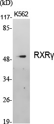
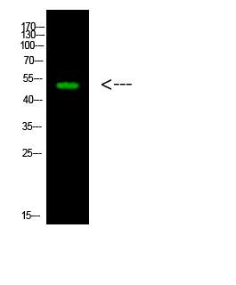
- Western Blot analysis of various cells using RXRγ Polyclonal Antibody cells nucleus extracted by Minute TM Cytoplasmic and Nuclear Fractionation kit (SC-003,Inventbiotech,MN,USA).
.jpg)
- Western Blot analysis of HepG2 cells using RXRγ Polyclonal Antibody cells nucleus extracted by Minute TM Cytoplasmic and Nuclear Fractionation kit (SC-003,Inventbiotech,MN,USA).
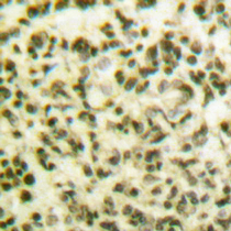
- Immunohistochemical analysis of paraffin-embedded Human lung cancer. Antibody was diluted at 1:100(4° overnight). High-pressure and temperature Tris-EDTA,pH8.0 was used for antigen retrieval.
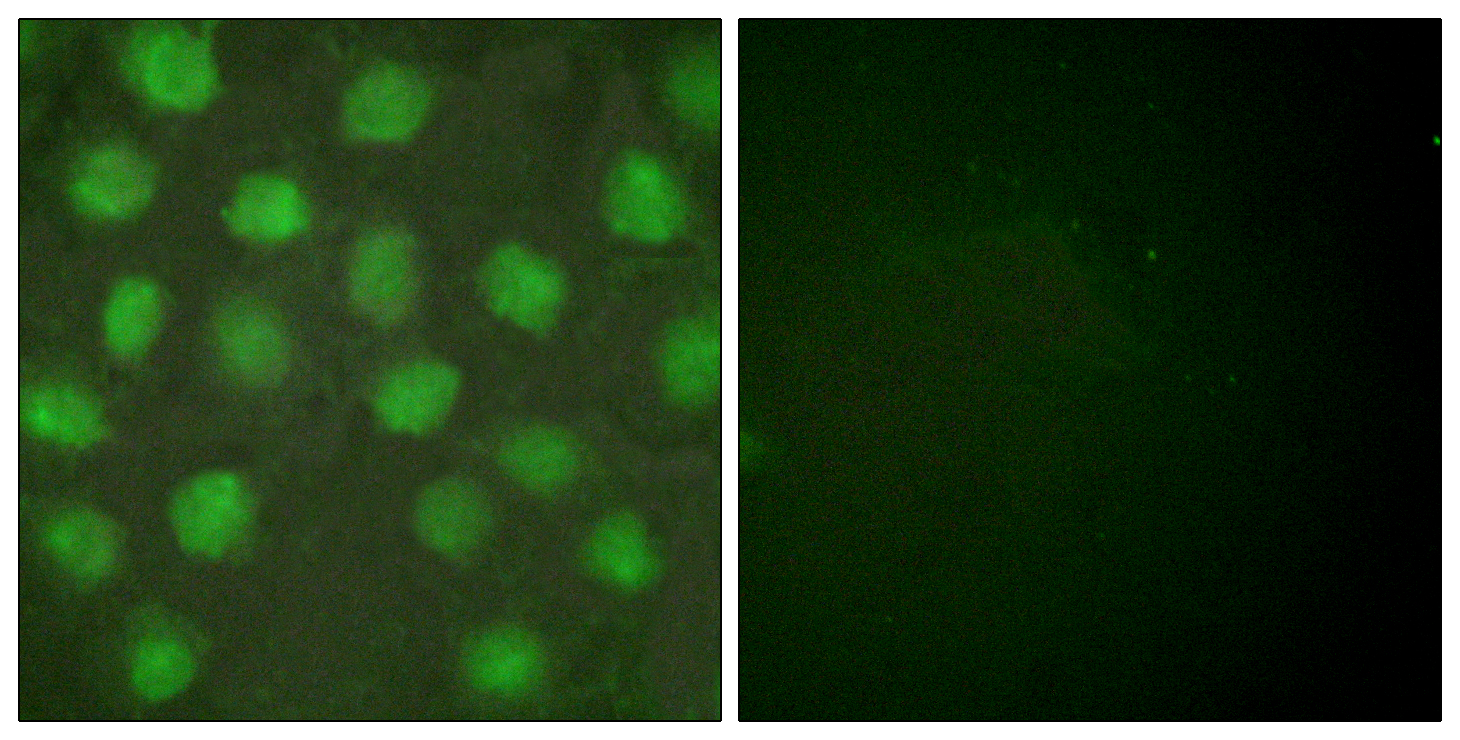
- Immunofluorescence analysis of HUVEC cells, using Retinoid X Receptor gamma Antibody. The picture on the right is blocked with the synthesized peptide.
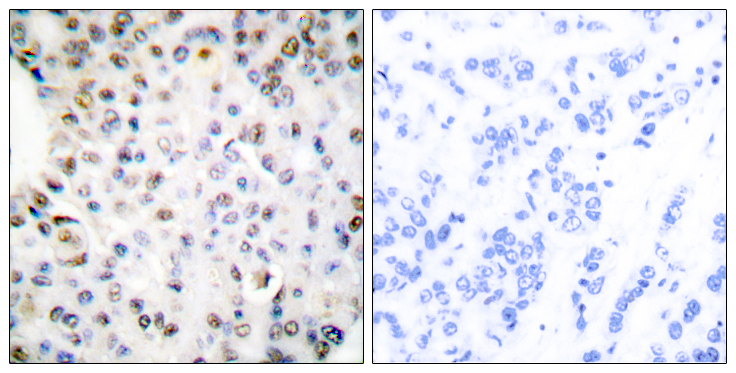
- Immunohistochemistry analysis of paraffin-embedded human breast carcinoma tissue, using Retinoid X Receptor gamma Antibody. The picture on the right is blocked with the synthesized peptide.
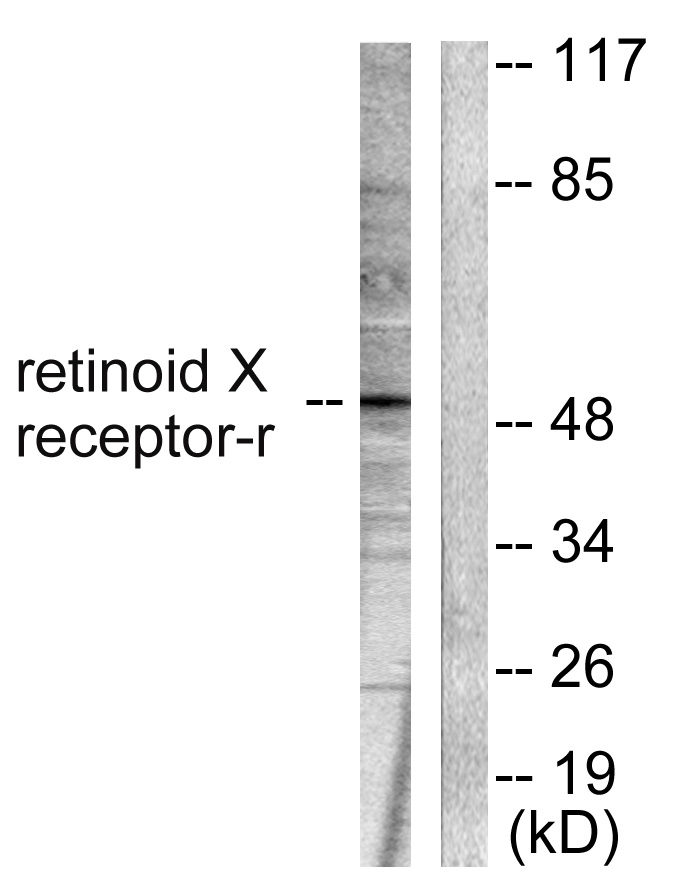
- Western blot analysis of lysates from HepG2 cells, using Retinoid X Receptor gamma Antibody. The lane on the right is blocked with the synthesized peptide.
