RGS10 Polyclonal Antibody
- Catalog No.:YT4071
- Applications:WB;IHC;IF;ELISA
- Reactivity:Human;Mouse;Rat
- Target:
- RGS10
- Gene Name:
- RGS10
- Protein Name:
- Regulator of G-protein signaling 10
- Human Gene Id:
- 6001
- Human Swiss Prot No:
- O43665
- Mouse Gene Id:
- 67865
- Mouse Swiss Prot No:
- Q9CQE5
- Rat Swiss Prot No:
- P49806
- Immunogen:
- The antiserum was produced against synthesized peptide derived from human RGS10. AA range:80-129
- Specificity:
- RGS10 Polyclonal Antibody detects endogenous levels of RGS10 protein.
- Formulation:
- Liquid in PBS containing 50% glycerol, 0.5% BSA and 0.02% sodium azide.
- Source:
- Polyclonal, Rabbit,IgG
- Dilution:
- WB 1:500 - 1:2000. IHC 1:100 - 1:300. ELISA: 1:10000.. IF 1:50-200
- Purification:
- The antibody was affinity-purified from rabbit antiserum by affinity-chromatography using epitope-specific immunogen.
- Concentration:
- 1 mg/ml
- Storage Stability:
- -15°C to -25°C/1 year(Do not lower than -25°C)
- Other Name:
- RGS10;Regulator of G-protein signaling 10;RGS10
- Observed Band(KD):
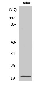
- 20kD
- Background:
- Regulator of G protein signaling (RGS) family members are regulatory molecules that act as GTPase activating proteins (GAPs) for G alpha subunits of heterotrimeric G proteins. RGS proteins are able to deactivate G protein subunits of the Gi alpha, Go alpha and Gq alpha subtypes. They drive G proteins into their inactive GDP-bound forms. Regulator of G protein signaling 10 belongs to this family. All RGS proteins share a conserved 120-amino acid sequence termed the RGS domain. This protein associates specifically with the activated forms of the two related G-protein subunits, G-alphai3 and G-alphaz but fails to interact with the structurally and functionally distinct G-alpha subunits. Regulator of G protein signaling 10 protein is localized in the nucleus. Two transcript variants encoding different isoforms have been found for this gene. [provided by RefSeq, Jul 2008],
- Function:
- function:Inhibits signal transduction by increasing the GTPase activity of G protein alpha subunits thereby driving them into their inactive GDP-bound form. Associates specifically with the activated forms of the G protein subunits G(i)-alpha and G(z)-alpha but fails to interact with the structurally and functionally distinct G(s)-alpha subunit. Activity on G(z)-alpha is inhibited by palmitoylation of the G-protein.,PTM:Isoform 3 is phosphorylated on Ser-16.,similarity:Contains 1 RGS domain.,
- Subcellular Location:
- [Isoform 1]: Cytoplasm, cytosol . Nucleus . Forskolin treatment promotes phosphorylation and translocation to the nucleus. .; Nucleus .
- Expression:
- Uterus,
- June 19-2018
- WESTERN IMMUNOBLOTTING PROTOCOL
- June 19-2018
- IMMUNOHISTOCHEMISTRY-PARAFFIN PROTOCOL
- June 19-2018
- IMMUNOFLUORESCENCE PROTOCOL
- September 08-2020
- FLOW-CYTOMEYRT-PROTOCOL
- May 20-2022
- Cell-Based ELISA│解您多样本WB检测之困扰
- July 13-2018
- CELL-BASED-ELISA-PROTOCOL-FOR-ACETYL-PROTEIN
- July 13-2018
- CELL-BASED-ELISA-PROTOCOL-FOR-PHOSPHO-PROTEIN
- July 13-2018
- Antibody-FAQs
- Products Images

- Western Blot analysis of various cells using RGS10 Polyclonal Antibody

- Western blot analysis of RGS10 Antibody. The lane on the right is blocked with the RGS10 peptide.

- Immunohistochemical analysis of paraffin-embedded human tonsil. 1, Antibody was diluted at 1:200(4° overnight). 2, Tris-EDTA,pH9.0 was used for antigen retrieval. 3,Secondary antibody was diluted at 1:200(room temperature, 45min).


