RELT Polyclonal Antibody
- Catalog No.:YT4046
- Applications:WB;IHC;IF;ELISA
- Reactivity:Human;Mouse
- Target:
- RELT
- Fields:
- >>Cytokine-cytokine receptor interaction
- Gene Name:
- RELT
- Protein Name:
- Tumor necrosis factor receptor superfamily member 19L
- Human Gene Id:
- 84957
- Human Swiss Prot No:
- Q969Z4
- Mouse Gene Id:
- 320100
- Mouse Swiss Prot No:
- Q8BX43
- Immunogen:
- The antiserum was produced against synthesized peptide derived from human RELT. AA range:381-430
- Specificity:
- RELT Polyclonal Antibody detects endogenous levels of RELT protein.
- Formulation:
- Liquid in PBS containing 50% glycerol, 0.5% BSA and 0.02% sodium azide.
- Source:
- Polyclonal, Rabbit,IgG
- Dilution:
- WB 1:500 - 1:2000. IHC 1:100 - 1:300. IF 1:200 - 1:1000. ELISA: 1:40000. Not yet tested in other applications.
- Purification:
- The antibody was affinity-purified from rabbit antiserum by affinity-chromatography using epitope-specific immunogen.
- Concentration:
- 1 mg/ml
- Storage Stability:
- -15°C to -25°C/1 year(Do not lower than -25°C)
- Other Name:
- RELT;TNFRSF19L;Tumor necrosis factor receptor superfamily member 19L;Receptor expressed in lymphoid tissues
- Observed Band(KD):
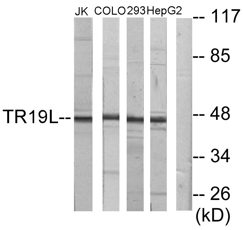
- 46kD
- Background:
- The protein encoded by this gene is a member of the TNF-receptor superfamily. This receptor is especially abundant in hematologic tissues. It has been shown to activate the NF-kappaB pathway and selectively bind TNF receptor-associated factor 1 (TRAF1). This receptor is capable of stimulating T-cell proliferation in the presence of CD3 signaling, which suggests its regulatory role in immune response. Two alternatively spliced transcript variants of this gene encoding the same protein have been reported. [provided by RefSeq, Jul 2008],
- Function:
- function:Mediates activation of NF-kappa-B. May play a role in T-cell activation.,PTM:Phosphorylated in vitro by OXSR1.,similarity:Belongs to the RELT family.,similarity:Contains 1 TNFR-Cys repeat.,subunit:Associates with TRAF1. Interacts with RELL1, RELL2 and OXSR1.,tissue specificity:Highest levels are in spleen, lymph node, thymus, peripheral blood leukocytes, bone marrow and fetal liver. Very low levels in skeletal muscle, testis and colon. Not detected in brain, kidney and pancreas.,
- Subcellular Location:
- Cell membrane ; Single-pass type I membrane protein . Cytoplasm . Cytoplasm, perinuclear region .
- Expression:
- Spleen, lymph node, brain, breast and peripheral blood leukocytes (at protein level) (PubMed:28688764). Expressed highly in bone marrow and fetal liver. Very low levels in skeletal muscle, testis and colon. Not detected in kidney and pancreas.
- June 19-2018
- WESTERN IMMUNOBLOTTING PROTOCOL
- June 19-2018
- IMMUNOHISTOCHEMISTRY-PARAFFIN PROTOCOL
- June 19-2018
- IMMUNOFLUORESCENCE PROTOCOL
- September 08-2020
- FLOW-CYTOMEYRT-PROTOCOL
- May 20-2022
- Cell-Based ELISA│解您多样本WB检测之困扰
- July 13-2018
- CELL-BASED-ELISA-PROTOCOL-FOR-ACETYL-PROTEIN
- July 13-2018
- CELL-BASED-ELISA-PROTOCOL-FOR-PHOSPHO-PROTEIN
- July 13-2018
- Antibody-FAQs
- Products Images

- Western blot analysis of lysates from Jurkat, COLO205, 293, and HepG2 cells, using RELT Antibody. The lane on the right is blocked with the synthesized peptide.

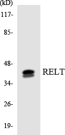
- Western blot analysis of the lysates from Jurkat cells using RELT antibody.

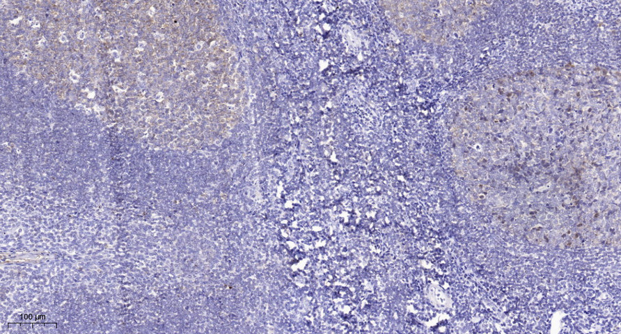
- Immunohistochemical analysis of paraffin-embedded human tonsil. 1, Tris-EDTA,pH9.0 was used for antigen retrieval. 2 Antibody was diluted at 1:200(4° overnight.3,Secondary antibody was diluted at 1:200(room temperature, 45min).


